Lambourile pe vase perforante tip “propeller” de la nivelul membrului inferior - aplicaåii clinice
Introducere: În reconstrucåiile membrului inferior au fost utilizate o mare varietate de lambouri începând cu lambourilepe circulaåie întâmplãtoare, lambourile axiale, transferul libermicrochirurgical şi multe variante ale acestora. Odatã cu aprofundarea cercetãrilor asupra vascularizaåiei cutanate şiîmbunãtãåirea tehnicilor microchirurgicale. lambourile pe vase perforante tip “propeller” au început sã fie studiate şi utilizate în clinicã.Material şi metodã: Un lambou tip “propeller” este definit ca unlambou insular mobilizat printr-o mişcare de rotaåie axialã înjurul unui pedicul vascular perforant pentru a acoperi defectul ce trebuie reconstruit. În concordanåã cu literatura despecialitate şi cu studiile efectuate la cadavru aceasta lucrareprezintã o clasificare a acestor lambouri în funcåie de mai multecriterii: 1) poziåia vasului perforant la nivelul lamboului; 2)forma insulei cutanate în funcåie de necesitãåile reconstructive;3) pediculul vascular al lamboului; 4) necesitãåile estetice şifuncåionale ale defectului de pãråi moi ce trebuie reconstruit.Bazându-ne pe datele din acest studiu am putut stabili indicaåiile specifice ale fiecarui tip de lambou. Concluzii: În strategia reconstructivã la nivelul membrului
inferior lambourile pe perforante tip “propeller” reprezintãprima opåiune pentru defectele de dimensiuni mici şi mediiîn anumite zone considerate “critice”. Cunoaşterea lungimiipediculului şi a suprafeåei lamboului permite alegerea optimã a acestuia pentru reconstrucåia unui anumit defecttopografic.
Cuvinte cheie: membru inferior, reconstrucåie, lambouri pevase perforante tip “propeller”, aplicaåii clinice
AbstractBackground: A great variety of flaps have been used in lowerlimb reconstructive surgery: random pattern flaps, axial flapsand free microsurgical transfers with many variants. Afterfurther research on the cutaneous blood supply and improve-ment of microsurgical techniques, perforator propeller flapsbegan to be studied and used.Material and method: A propeller flap is defined as an insularflap mobilized through an axial rotation on a perforator pediclein order to cover a defect that has to be reconstructed.According to the specialized literature data and to the studieswe made on cadavers, this paper establishes a classificationbased on several criteria: 1) the position of the supplying perforator; 2) the reconstructive necessities and the skin island;3) the flap blood supply; 4) the aesthetic and functionaldemands of the reconstructed areas. Based on this study weestablished the specific reconstructive indications for these propeller flaps.Conclusions: The reconstructive lower limb strategy allowsthe use of the perforator propeller flaps as first therapeuticoption for small and medium defects in certain areas
1Plastic Surgery Department, “Sfanta Maria” Clinical Hospital, Bucharest, Romania2“Carol Davila” University of Medicine and Pharmacy, Plastic Surgery and Reconstructive Microsurgery Clinic, Bucharest Clinical Emergency Hospital, Romania
considered as “critical”. Knowledge of the pedicle lengthand the surface of the flaps facilitates the optimum flapchoice in the reconstruction of a certain area.
Complex wounds need complex reconstructions and this wasnot possible until different types of flaps were developed. Theimportant contribution of describing the fascial and muscle toskin circulation through septocutaneous and musculocutaneousvessels was the first step in the evolution of flaps and led to an“explosion” in the use of musculocutaneous flaps for recon-struction (1). However, the impact of using fasciocutaneousflaps was minor compared to that of musculocutaneous flaps,but also led to a more detailed research of the skin vascular supply. Thus, the angiosome concept was born and, based oncadaveric injection studies, the vascular territories of the bodywere described (there were 21 angiosomes in the lower extremity) (2).The source vessels for the skin of the thigh arethe superficial and profunda femoris arteries through their musculocutaneous and septocutaneous branches with 30 to 50skin perforators. In the popliteal fossa, the superficial femoralartery becomes the popliteal artery which gives birth to thethree main vascular sources of the leg: the anterior and posterior tibial arteries and the peroneal artery with their corresponding muscolocutaneous and septocutaneous perfora-tors (there are 20 to 40 perforators). In the foot region, the posterior tibial artery branches in medial and lateral plantararteries, the anterior tibial artery becomes the dorsalis pedisartery and the peroneal artery ends as the lateral calcanealartery. There are multiple anastomoses between them thatmake it difficult to define vascular territories at this level. Thesevessels are the origin of small musculocutaneous and septo-cutaneous perforators (2).
The last 20 years of reconstructive surgery developmentwere marked by the appearance of some new flaps based on perforator blood vessels. In 1989 Koshima and Soeda used forthe first time the term “perforator flap” for a paraumbilical skinflap based on a muscular perforator. The following researchescarried out especially in the Asian schools have led to theappearance and description of numerous flaps based on perforator blood vessels and implicitly to the appearance ofnumerous confusions and controversies. The Gent Conventionclarified and standardized the terminology and classification ofperforator flaps (3).
The refinement of the surgical procedures and the researchconcerning the perforator blood vessels have been followed bythe discovery of a new type of perforator flap - the “propeller”type flap. The term was introduced by Hyakusoku in 1991 inorder to describe an insular skin flap based on a randomized
subcutaneous pedicle which may be rotated 90 degrees aroundthe pedicle similar to a propeller’s rotation. Hallock uses thesame name for a flap like the one described by Hyakusoku, butthis time the flap was based on a skeletonized perforator vessel.The growing interest for these flaps and the clinical applica-tions in reconstructing soft tissues losses have imposed thestandardization of their definition and terminology at theTokyo convention in 2009.
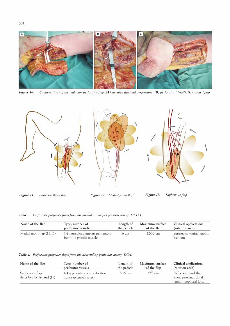
A propeller flap is now defined as an insular flap mobilizedthrough an axial rotation in order to cover the reconstructeddefect, thus excluding from the definition of such a flapadvanced insular flaps, type V-Y flaps or rotated peninsularflaps. The perforator propeller flap is a propeller type flapwhose skin island is centred on a perforator vessel which corresponds to the pivot point of the flap. The two portions ofthe skin island (the blades or paddles of the propeller) may beequal or unequal and they may rotate around the pedicleaccording to the reconstructive necessities between 90-180degrees, similar to a propeller’s movement. Even if the difference in dimensions and forms between the paddles of theskin island makes the resemblance to a propeller less obvious,such a flap is not excluded from the definition. The rotationdegree and the dimensions of the two portions of the skinisland of the propeller flap are determined by the position ofthe accommodated perforator flap to the tissue defect thatmust be reconstructed. The 180 degrees rotation might beobtained if there is enough tissue that can be mobilizedthrough axial rotation in the extension of the axis connectingthe defect with the pivot point of the perforator vessel. Thecloser the position of the perforator vessel to the defect is, themore will the dimensions of the two paddles of the skin islandbecome unequal (4,5,6,7).
Classification according to the position of the supplyingperforator vessel, the reconstructive necessities and the formof the skin island used for reconstruction, the flaps’ bloodsupply, the aesthetic and functional demands of the defect(8,9,10):
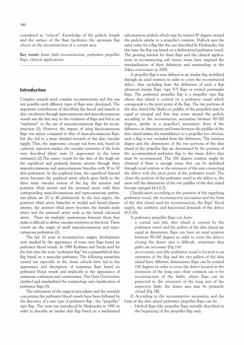
1) perforator propeller flaps can have: - a central axis (the skin island is centred by the
perforator vessel and the pallets of the skin island areequal in dimension; flaps can have an axial rotationbetween 90-180 degrees in order to cover the defect;closing the donor area is difficult, sometimes skingrafts are necessary (Fig.1A)
- an eccentric axis (the perforator vessel is located to anextremity of the flap and the two pallets of the skinisland have different dimensions; flaps can be rotated180 degrees in order to cover the defect located in theextension of the long axis; their common use is forreconstruction of the limbs, where flaps can be projected in the extension of the long axis of therespective limb; the donor area may be primarilyclosed (Fig.1B).
2) According to the reconstructive necessities and theform of the skin island perforator propeller flaps can be:
- bilobed flaps (the propeller flaps initially described inthe beginning of the propeller flap use);
300
- polilobed flaps, indicated especially for scar contrac-ture release;
- hockey stick flaps indicated for the optimal coverage ofsome defects and avoidance at the same time of somephysical obstacles (external fixtures) or anatomical areas(scars);
- “free style” flaps – such flaps imply an identificationof the pivot point and of the flap’s rotation arc; offersfreedom in modification of the form and dimensionsof the skin island according to the defect during surgery.
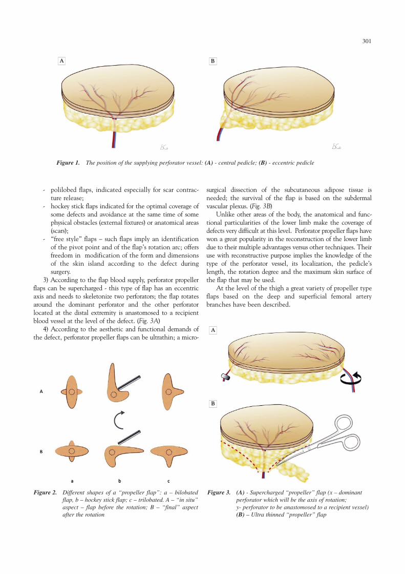
3) According to the flap blood supply, perforator propellerflaps can be supercharged - this type of flap has an eccentricaxis and needs to skeletonize two perforators; the flap rotatesaround the dominant perforator and the other perforator located at the distal extremity is anastomosed to a recipientblood vessel at the level of the defect. (Fig. 3A)
4) According to the aesthetic and functional demands ofthe defect, perforator propeller flaps can be ultrathin; a micro-
surgical dissection of the subcutaneous adipose tissue is needed; the survival of the flap is based on the subdermal vascular plexus. (Fig. 3B)
Unlike other areas of the body, the anatomical and func-tional particularities of the lower limb make the coverage ofdefects very difficult at this level. Perforator propeller flaps havewon a great popularity in the reconstruction of the lower limbdue to their multiple advantages versus other techniques. Theiruse with reconstructive purpose implies the knowledge of thetype of the perforator vessel, its localization, the pedicle’slength, the rotation degree and the maximum skin surface ofthe flap that may be used.
At the level of the thigh a great variety of propeller typeflaps based on the deep and superficial femoral arterybranches have been described.
Figure 1. The position of the supplying perforator vessel: (A) - central pedicle; (B) - eccentric pedicle
AA BB
Figure 2. Different shapes of a “propeller flap”: a – bilobatedflap, b – hockey stick flap; c – trilobated. A – “in situ”aspect – flap before the rotation; B – “final” aspectafter the rotation
Figure 3. (A) - Supercharged “propeller” flap (x – dominant perforator which will be the axis of rotation; y- perforator to be anastomosed to a recipient vessel)(B) – Ultra thinned “propeller” flap
AA
BB
301
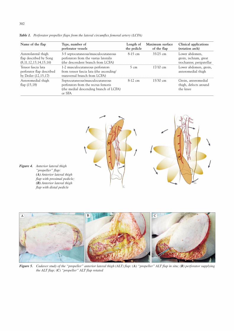
Table 1. Perforator propeller flaps from the lateral circumflex femoral artery (LCFA)
Name of the flap Type, number of Length of Maximum surface Clinical applicationsperforator vessels the pedicle of the flap (rotation arch)
Anterolateral thigh 3-5 septocutaneous/musculocutaneous 8-15 cm 35/25 cm Lower abdomen,flap described by Song perforators from the vastus lateralis groin, ischium, great(8,11,12,13,14,15,16) (the descendent branch from LCFA) trochanter, peripatellar Tensor fascia lata 1-2 musculocutaneous perforators 5 cm 17/10 cm Lower abdomen, groin,perforator flap described from tensor fascia lata (the ascending/ anteromedial thighby Deiler (12,15,17) transversal branch from LCFA) Anteromedial thigh Septocutaneous/musculocutaneous 8-12 cm 15/30 cm Groin, anteromedial flap (15,18) perforators from the rectus femoris thigh, defects around
(the medial descending branch of LCFA) the kneeor SFA
Figure 4. Anterior lateral thigh “propeller” flap: (A) Anterior lateral thighflap with proximal pedicle;(B) Anterior lateral thighflap with distal pedicle
Figure 5. Cadaver study of the “propeller” anterior lateral thigh (ALT) flap: (A) “propeller” ALT flap in situ; (B) perforator supplyingthe ALT flap; (C) “propeller” ALT flap rotated
AA BB CC
302
Table 2. Perforator propeller flaps from the profunda femoris artery (PFA)
Name of the flap Type, number of Length of Maximum surface Clinical applicationsperforator vessels the pedicle of the flap (rotation arch)
Lateral thigh flap (15,19) The second septocutaneous 5-7 cm 8/18 cm great trochanter, ischiumperforator from PFA or musculocutaneous perforators from the biceps femoris
Adductor perforator First musculocutaneous 8-9 cm 25/30 cm Great trochanter, flap (15,17,19) perforator from adductor magnus ischium, perineal defects
(first medial perforator of PFA) Posterior thigh flap The third septocutaneous 5-6 cm Popliteal fossa, defectsdescribed by Song (15,19) perforator from PFA around the knee
Figure 6. Tensor fascia lata perforator flap: (A) front view; (B) lateral view Figure 7. Anterior medial thigh perforator flap
Table 3. Perforator propeller flaps from the medial circumflex femoral artery (MCFA)
Name of the flap Type, number of Length of Maximum surface Clinical applicationsperforator vessels the pedicle of the flap (rotation arch)
Medial groin flap (15,17) 1-2 musculocutaneous perforators 6 cm 12/30 cm perineum, vagina, groin,from the gracilis muscle ischium
Table 4. Perforator propeller flaps from the descending genicular artery (DGA)
Name of the flap Type, number of Length of Maximum surface Clinical applicationsperforator vessels the pedicle of the flap (rotation arch)
Saphenous flap 1-4 septocutaneous perforators 5-15 cm 29/8 cm Defects around thedescribed by Acland (15) from saphenous artery knee, proximal tibial
region, popliteal fossa
304
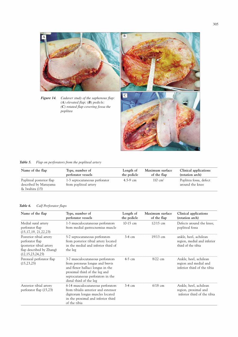
Table 5. Flap on perforators from the popliteal artery
Name of the flap Type, number of Length of Maximum surface Clinical applicationsperforator vessels the pedicle of the flap (rotation arch)
Popliteal posterior flap 1-3 septocutaneous perforator 4.5-9 cm 110 cm2 Poplitea fossa, defectdescribed by Maruyama from popliteal artery around the knee& Iwahira (15)
Figure 14. Cadaver study of the saphenous flap: (A) elevated flap; (B) pedicle; (C) rotated flap covering fossa thepoplitea
AA BB
CC
Table 6. Calf Perforator flaps
Name of the flap Type, number of Length of Maximum surface Clinical applicationsperforator vessels the pedicle of the flap (rotation arch)
Medial sural artery 1-3 musculocutaneous perforators 10-15 cm 12/15 cm Defects around the knee,perforator flap from medial gastrocnemius muscle popliteal fossa(15,17,19, 21,22,23)Posterior tibial artery 5-7 septocutaneous perforators 3-4 cm 19/13 cm ankle, heel, achileanperforator flap from posterior tibial artery located region, medial and inferior(posterior tibial artery in the medial and inferior third of third of the tibiaflap described by Zhang) the leg(12,15,23,24,25)Peroneal perforator flap 3-7 musculocutaneous perforators 4-5 cm 8/22 cm Ankle, heel, achilean(15,23,25) from peroneus longus and brevis region and medial and
and flexor halluci longus in the inferior third of the tibiaproximal third of the leg and septocutaneous perforators in the distal third of the leg
Anterior tibial artery 6-14 musculocutaneous perforators 3-4 cm 6/18 cm Ankle, heel, achileanperforator flap (15,23) from tibialis anterior and extensor region, proximal and
digitorum longus muscles located inferior third of the tibiain the proximal and inferior third of the tibia
305
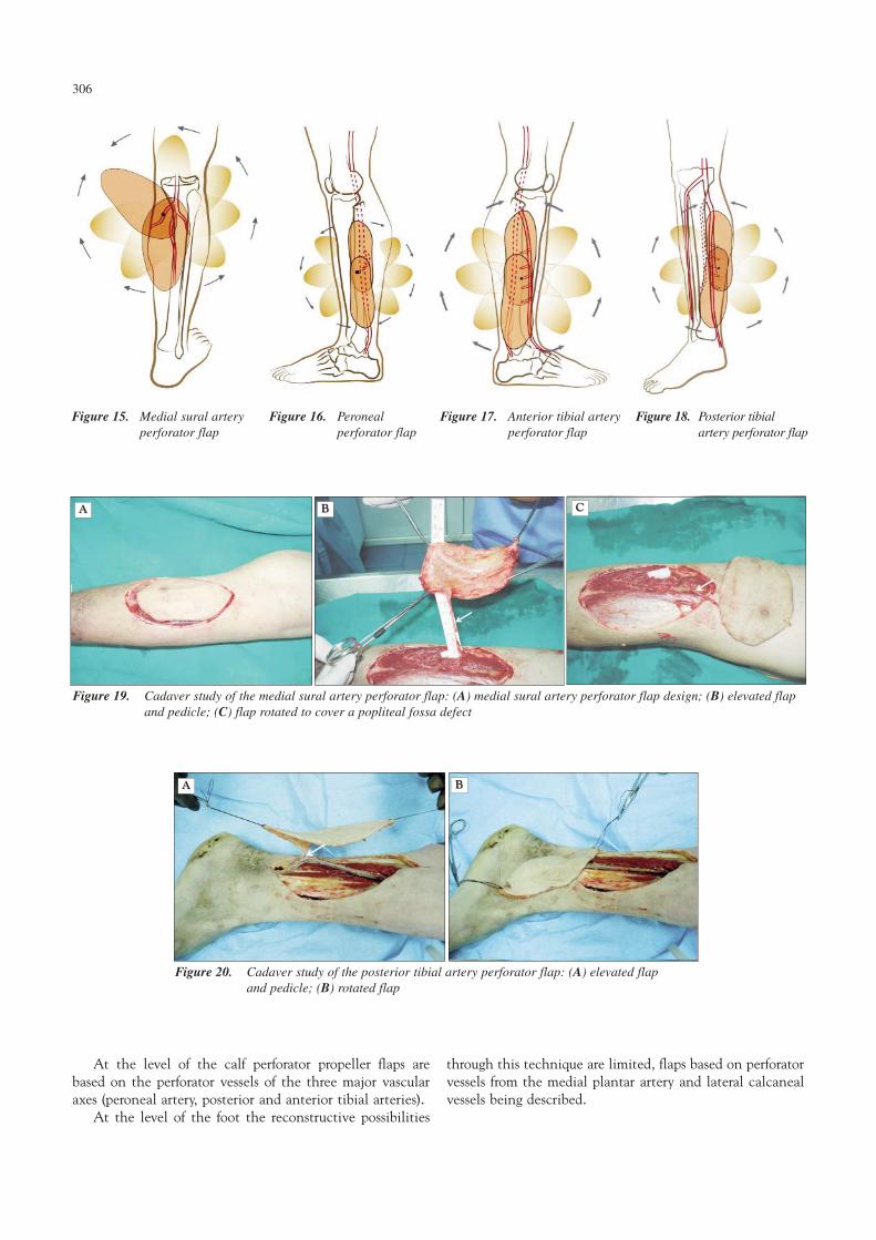
Figure 15. Medial sural arteryperforator flap
Figure 16. Peroneal perforator flap
Figure 17. Anterior tibial arteryperforator flap
Figure 18. Posterior tibialartery perforator flap
Figure 19. Cadaver study of the medial sural artery perforator flap: (A) medial sural artery perforator flap design; (B) elevated flapand pedicle; (C) flap rotated to cover a popliteal fossa defect
AA BB CC
Figure 20. Cadaver study of the posterior tibial artery perforator flap: (A) elevated flapand pedicle; (B) rotated flap
AA BB
At the level of the calf perforator propeller flaps arebased on the perforator vessels of the three major vascularaxes (peroneal artery, posterior and anterior tibial arteries).
At the level of the foot the reconstructive possibilities
through this technique are limited, flaps based on perforatorvessels from the medial plantar artery and lateral calcanealvessels being described.
306
Figure 21. Clinical case using a posterior tibial artery perforator flap: (A) soft tissuedefect to cover; (B) elevated flap and perforators; (C) rotated flap on the proximal perforator pedicle; (D) flap rotated in final position
AA BB CC
DD
Table 7. Foot perforator flaps
Name of the flap Type, number of Length of Maximum surface Clinical applicationsperforator vessels the pedicle of the flap (rotation arch)
Lateral calcaneal direct perforator from lateral - - Achilean region, internal artery perforator flap (15) calcaneal artery malleolus, heelMedial plantar artery septocutaneous perforator from 2-3 cm 4/8 cm Heel, achilean region,perforator flap (15,26) medial plantar artery internal and external
Reconstructive surgery of the lower limb level represents a fieldin continuous development in which new strategies and newsurgical techniques are permanently implemented. Perforatorpropeller flaps have won fast their popularity due to their multiple advantages versus other techniques.
These advantages of the perforator flaps are (9,27,28,29): - presence in the same operating area with the defect to
be reconstructed, - reduction of the operating time, - possibility to cover the defect with tissues having
similar qualities, - these flaps do not sacrifice deep musculature (they
conserve the function), - flaps can be raised together with other structures
(fascia, nerve, muscle) for complex reconstructions, - it is not needed to sacrifice the main vascular axis, - perforator flaps don’t require microsurgical anastomosis, - these flaps allow to reduce the morbidity of the donor
area (the axial rotation movement allows its partialcoverage with the flap),
- a better aesthetic appearance can be achieved (theycan be thinned through microsurgical procedures inorder to cover joint surfaces),
- reconstruction of soft tissue defects with these flaps canbe used also on patients with peripheral obstructiveartery disease after revascularization,
- the free-style principle allows the adjustment of theflap’s form and dimensions during surgery (12).
Perforator propeller flaps present also a series of disadvan-tages (30):
- multiple anatomical variants of the perforator vessels,- these flaps require the preoperative identification of
the perforator vessels, - microsurgical abilities of the surgeon are needed,- these flaps cannot cover defects with large surface
(except for the flap on LCFA perforator).The reconstructive strategy of the lower limb allows the
use of perforator propeller flaps as first therapeutic option forthe defects of small and medium dimensions in certain areasconsidered “critical” from the reconstructive point of view.
For trochanterian and ischiatic defects one can use perforator propeller flaps from:
- LCFA (anterolateral thigh flap)- PFA (posterolateral thigh flap, adductor flap)- MCFA (medial groin flap)The defects from the groin area can be covered with
- Popliteal artery (popliteoposterior flap)- Medial sural arteryFor the defects from the popliteal fossa perforator
propeller flaps can be raised from: - DGA (saphenous flap)- Popliteal artery (popliteoposterior flap)- Medial sural arteryThe defects from the achilean area, heels and ankle can
Over the last years, perforator propeller flaps have enlarged thearea of therapeutic options for the reconstruction of soft tissuedefects at the lower limb level even becoming the first choicein case of soft tissue losses of small or medium dimensions,especially at the level of the lower leg and foot (24,31). In ourcountry, among the variety of flaps and microsurgical procedures that are used for lower limb reconstruction (32,34)there is also an initial experience in using perforator propellerflaps for lower limb reconstruction in patients with peripheralarterial obstructive disease (34).
Authors contribution
All authors had equal scientific contributions.
Acknowledgement
This work is partially supported by the OperationalProgramme Human Resources Development (HRD),financed by the European Social Fund and the RomanianGovernment Decision no. POSDRU/89/1.5/S/64153
The dissection study was conducted with the permissionof the Ethics Committee of the Forensic Medicine InstituteBucharest.
ReferencesReferences
1. Mathes SJ, Nahai F. Reconstructive surgery: principles, anatomy,& technique. The reconstructive triangle: a paradigm for surgical decision making, Volume 1. New York: ChurchillLivingstone; 1997.
2. Blondeel PN, Morris SF, Hallock GG, Nelligan PC. VascularAnatomy of the Integument. Perforator Flaps vol 1.
3. Blondeel PN, Van Landuyt KHI, Monstrey SJM, Hamdi M,Matton GE, Allen RJ, Duppin C, Feller AM, Koshima I,Kostadoglu N, Wei FC. The “Gent” Consensus on PerforatorFlap Terminology: Preliminary Definition. Plastic andReconstructive Surgery. 2003;112(5):1378-82.
4. Pignatti M, D’Arpa S, Cubison TCS. Novel FasciocutaneousFlaps for the Reconstruction of Complicated Lower Extremity
308
Wounds. Techniques in Orthopaedics. 2009;24(2):88-95.5. Pignati M, Ogava R, Hallock GG, Mateev M, Georgescu Al,
Balakrishanan G, et al. The "Tokyo" consensus on propellerflaps. Plast Reconstr Surg. 2011;127(2):716-22.
6. Teo TC. The propeller Flap Concept. Clinics in PlasticSurgery. 2010;37(4):615-26.
7. Demirtas Y, Ozturk N, Kelahmetoglu O, Demir A. PedicledPerforator Flaps. Techniques in Knee Surgery. 2009;8(2):119-25.
8. Ayestaray B, Ogawa R, Hyakusoku H. Propeller flaps: classifi-cations and clinical applications. Ann Chir Plast Esthet. 2011;56(2):90-8.
9. Hiakusoku H, Ogawa R, Oky K, Ishii N. The perforator pedicled propeller (PPP) flap method: report of two cases. JNippon Med Sch. 2007;74(5):367-71.
10. Murakami M, Hyakusoku H, Ogawa R. The MultilobedPropeller Flap Method. Plastic and Reconstructive Surgery.2005;116(2):599-604.
11. Chen CY, Hsieh CH, Kuo YR, Jeng SF. An AnterolateralThigh Perforator Flap from the Ipsilateral Thigh for Soft-Tissue Reconstruction around the Knee. Plastic andReconstructive Surgery. 2007;120(2):470-473.
12. Wei FC, Mardini S. Flaps and Reconstructive surgery.Anterolateral Thigh Flap pg.539-558, Tensor fascia Lata Flappg. 559-574, Posterior Tibial Artery Perforator Flap pg. 601-617, Free Style Flap pg. 617-624.
13. Park J, Rodrigues E, Bluebond-Langer R, Bochicchino G,Cristy MR, Bochicchino K, Scalea TM. The AnterolateralThigh Flap is Highly Effective for Reconstruction of ComplexLower Extremity Trauma. The Journal of Trauma Injury,Infection and Critical Care. 2007.
14. Nelligan PC, Lannon DA. Versatility of the PedicledAnterolateral Thigh Flap. Clinics in Plastic Surgery. 2010;37(4):677-82.
20. Thione A, Valdatta L, Buoro M, Tuinder S, Mortarini C, PutzR. The medial sural artery perforators: anatomic basis for a surgical plan. Ann Plast Surg. 2004;53(3):250-5.
21. Hallock GG. The medial sural(MEDIAL GASTROCNEMIUS)perforator local flap. Ann Plast Surg. 2004;53(5):501-5.
22. Kim HH, Jeong JH, Seul JH, Cho BC. New design and iden-tification of the medial sural perforator flap: an anatomicalstudy and its clinical applications. Plast Reconstr Surg.2006;117(5):1609-18.
23. Schaverien M, Saint-Cyr M. Perforators of the lower leg:analysis of perforator locations and clinical application forpedicled perforator flaps. Plast Reconstr Surg. 2008;122(1):161-70.
24. Masia J, Moscatiello F, Pons G, Fernandez M, Lopez S, SerretP. Our Experience in Lower limb Reconstruction withPerforator Flaps. Annals of Plastic Surgery. 2007;58(5):507-12.
25. Jakubietz R, Jakubietz MG, Gruenert JG, Kloss DF. The 180-degree perforator-based propeller flap for soft tissue coverageof the distal, lower extremity: a new method to achieve reliablecoverage of the distal lower extremity with a local, fasciocuta-neous perforator flap. Ann Plast Surg. 2007;59(6):667-71.
26. Koshima I, Narushima M, Mihara M, Nakai I, Akazawa S,Fukuda N, et al. Island medial plantar artery perforator flap forreconstruction of plantar defects. Ann Plast Surg. 2007;59(5):558-62.
27. Geddes CR, Morris SF, Neligan PC. Perforator flaps: evolution,classification, and applications. Ann Plast Surg. 2003;50(1):90-9.
28. Hallock GG. Lower Extremity Muscle Perforator Flaps for LowerExtremity Reconstruction. Pl. and Rec. Surg. 2004;114(5):1123-29.
29. Saint-Cyr M, Scaverien MV, Rorich RJ. Perforator Flaps:Phisiology, Anatomy and Use in Reconstruction. Pl. and Rec.Surg. 2009;123(4):132-45.
30. Sing DP, Brewer MB, Silverman RP. Reconstruction of LowerExtremity Wounds Using Perforator Flaps. Techniques inOrthopaedics. 2009;24(2):123-29.
31. Herman C, Hoschander AS, Strauch B. New Strategies inSurgical Reconstruction of the Lower Extremity. Techniques inOrthopaedics. 2009;24(2):123-29.
32. Iliescu V, Jiga LP, Ionac M. Complex microsurgical reconstruc-tion of diabetic foot. Chirurgia (Bucur). 2005;100(2):181-6.Romanian
33. Jecan CR, Alecu L, Costan IÆ. Comparative study of threeseries of neuro-cutaneous flaps used for coverage of leg defects.Chirurgia (Bucur). 2011;106(6):781-8. Romanian
34 . Jiga LP, Barac S, Taranu G, Blidisel A, Dornean V, Nistor A,et al. The versatility of propeller flaps for lower limb recons-truction in patients with peripheral arterial obstructive disease: initial experience. Ann Plast Surg. 2010;64(2):193-7.