Submission: Only original papers in English are considered and should be sent to the following address: [email protected]
Manuscripts should be submitted by e-mail only, written in Microsoft Word 97 or later versions.
Conditions: AII manuscripts are subject to editorial review. Manuscripts are received with the explicit understanding that they are not under simultaneous consideration by any other publication. Submission of an article for publication implies the transfer of the Copyright from the author the publisher upon acceptance. Accepted papers become the permanent property of "Fiziologia" (Physiology) and may not be reproduced by any means, in-whole or in part, without the written consent of the publisher. It is the author's responsibility to obtain permission to reproduce illustrations, tables, etc. from other publications.
Arrangement: Title page: The first of each paper should indicate
the title, the authors' names and their affiliation(s). A short title for use as running head is also required.
Keywords: for indexing purposes, a list of 3-5 keywords in English and Romanian is essential.
Corresponding author: Indicate the full name, the email address and the phone number.
Abstract: Each paper needs abstract and title in Romanian and English language, fonts size 9, Arial Narrow.
Body text: fonts size 10, Arial Narrow. Small type: Paragraphs which can or must be set
in smaller type (case histories, test methods, etc.) should be indicated with a „p" (petit) in the margin on the left-hand side.
Footnotes: Avoid footnotes. When essential, they are numbered consecutively and typed at the foot of the appropriate page, fonts size 8, Arial Narrow.
Tables and illustrations: Tables (numbered in Roman numerals) and illustrations (numbered in Arabic numerals) should be prepared on separate sheets, fonts size 9, Arial Narrow. Tables require a heading, and figures a legend, also prepared on a separate sheet. For the reproduction of illustrations, only good drawings and original photographs can be accepted; negatives or photocopies cannot be used. When possible, group several illustrations on one block for reproduction (max. size 140x188 mm) or
provide crop marks. On the back of each illustration indicate its number, the author's name, and article title.
References: In the text identify references by Arabic figures, (in brackets), fonts size 9, Arial Narrow. Material submitted for publication but not yet accepted should be noted as "unpublished data" and not be included in the reference list. The list of references should include only those publications which are cited in the text. The references should be numbered and arranged alphabetically by the authors' names. The surnames of the authors followed by initials should be given. There should be no punctuation signs other than a comma to separate the authors. When there are more than 3 authors, the names of the 3 only are used, followed by "et al" abbreviate journal names according to the Index Medicus system. (also see International Committee of Medical Journal Editors: Uniform Requirements for manuscripts submitted to biomedical journals. Ann Intern Med 1982; 96: 766-771).
Examples: (a) Papers published in periodicals: Kauffman
HF, van der Heide S, Beaumont F, et al: Class-specific
antibody determination against Aspergillus fumigatus
by mean of the enzyme-linked immunosorbent assay.
III. Comparative study: IgG, IgA, IgM, ELISA titers,
precipitating antibodies and IGE biding after
fractionation of the antigen. Int Arch Allergy Appl
Immunol 1986; 80:300 - 306.
(b) Monographs; Matthews DE, Farewell VT:
Using and Understanding Medical Statistics. Basel, Karger,
1985.
(c) Edited books: Hardy WD Jr, Essex M:.FeLV-
inducted feline acquired immune deficiency syndrome: A
model for human AIDS; in Klein E(ed): Acquired
Immunodeficiency Syndrome. Prag Allergy, Busel,
Karger, 1986, vol 37,353 - 376.
Galley proofs: unless indicated otherwise, galley proofs are sent to the first-named author and should be returned with the least possible delay. Alternations made in galley proofs, other than the corrections of printer's errors, are charged to the author. No page proofs are supplied.
Fiziologia - Physiology • 2017.27.1(93) 3
CONTENTS
1. Clinico-Pathological Evaluation of Pregnancy Induced Hypertension ................................................. 4
Zaha DC, Vesa C, Cismas-Pruteanu P, Daina LG
2. Hyperuricemia – The New Cardiovascular Risk Factor - Consequences and Effects ......................... 9
Moisi MI, Vesa CM, Ardelean AI, Zaha D, Cismaş-Pruteanu P, Magyar I, Popescu MI
3. Implication of Distress and Emotional Intelligence in Forensic Practice ........................................... 14
Cojocaru A, Ageu L
4. Associated Congenital Anomalies in New-Borns Diagnosed with Congenital
Diaphragmatic Hernia ............................................................................................................................ 17 Vesa C, Zaha D, Cismas-Pruteanu P, Moisi M, Mekeres F, Jurca C
5. Depressive Pathology and Bipolar Disorder: Particularities of the Theory of Mind .......................... 20
Homorogan C, Barboianu R, Rivis I, Tulbure BT, Giurgi-Oncu C, Bredicean C
6. Pulmonary Aspergilloma in an Immunocompetent Patient. A Case Report ....................................... 24
Cioboata R, Bazavan I, Georgescu M, Chaudrey S, Gaman AE
CUPRINS
1. Evaluarea clinico-patologică a hipertensiunii induse de sarcină .......................................................... 4 Zaha DC, Vesa C, Pruteanu Cismas P, Daina LG
2. Hiperuricemia asimptomatica - noul factor de risc cardiovascular? - Consecințe și efecte .............. 9
Moisi MI, Vesa CM, Ardelean AI, Zaha D, Cismaş Pruteanu P, Magyar I, Popescu MI
3. Implicațiile distresului și ale inteligenței emoționale în activitatea medicilor legiști ........................ 14
Cojocaru A, Ageu L
4. Anomalii congenitale asociate la nou-născuții cu hernie diafragmatică congenitală ........................ 17
Vesa C, Zaha D, Cismas-Pruteanu P, Moisi M, Mekeres F, Jurca C
5. Patologia depresivă și tulburările bipolare: particularități ale teoriei minții ....................................... 20
Homorogan C, Barboianu R, Rivis I, Tulbure BT, Giurgi-Oncu C, Bredicean C
6. Aspergilom pulmonar la un pacient immunocompetent. Prezentare de caz ...................................... 24
Cioboata R, Bazavan I, Georgescu M, Chaudrey S, Gaman AE
4 Fiziologia - Physiology • 2017.27.1(93)
CLINICO-PATHOLOGICAL EVALUATION
OF PREGNANCY INDUCED HYPERTENSION
DANA CARMEN ZAHA, COSMIN VESA, PETRISOR PRUTEANU CISMAS,
LUCIA GEORGETA DAINA
University of Oradea, Faculty of Medicine and Pharmacy of Oradea
ABSTRACT Introduction. ecause the clinical manifestation of preeclampsia can be heterogeneous, laboratory tests can be very useful for early preeclampsia diagnosis in pregnancy induced hypertension patients. Method. A total 110 patients with clinical diagnosis of pregnancy induced hypertension (PIH) were included in the study. Complete blood count, urine examination, biochemical tests (ALT, AST, LDH, T-bilirubin, uric acid) and coagulation tests were done in all patients. The urinary test checked the usual parameters and proteinuria (24-hour urine collection). In addition, protein-creatinine ratio was evaluated in urinary specimen with a rapid test Results. In our study, 66 patients had mild PIH and 44 severe PIH. Headache was the predominant symptom followed by giddiness. Pathological changes of the biochemical, hematological and coagulation parameters were observed mainly in severe PIH. Conclusion. Patients significantly younger, low parity had significantly increased systolic and diastolic pressure and changes of values of liver enzymes, uric acid, urine protein, and LDH levels. Abnormalities (qualitative and quantitative abnormalities) of red blood cells and platelets are frequently seen in PIH. Spot urinary P/C ratio in pregnancy induced hypertension patients can be used as a screening test as a good predictor for significant proteinuria and could not be used for diagnostic tests to replace 24-h urine protein measurements. Keywords: pregnancy, hypertension, preeclampsia, diagnostic.
INTRODUCTION
Pregnancy-induced hypertension (PIH) remains a major cause of perinatal and maternal morbidity and mortality, occurring in 6-10% of all pregnancies [1]. It is defined as systolic blood pressure (SBP) >140 mmHg and diastolic blood pressure (DBP) >90 mmHg and classified as mild (SBP 140-149 and DBP 90-99 mmHg), moderate (SBP 150-159 and DBP 100-109 mmHg) and severe (SBP ≥160 and DBP ≥110 mmHg).
To a certain extent, mothers suffer from major compli-cations like development of disseminated intravascular coagulation (DIC), cerebrovascular accidents, renal failure, retinal detachment, pulmonary edema, liver rupture, abruptio placentae, and death. In the case of the fetus, intrauterine growth retardation, prematurity and intrauterine death may occur, all being the consequences of placental insufficiency. Preeclampsia is characterized by the appearance of hypertension and proteinuria, usually after 20 weeks of gestation. There are many tests or methods reported in the literature for predicting the development of preeclampsia, but none of them had shown sufficient predictive value.
HELLP syndrome, a severe form of pre-eclampsia develops in 4-12% of women with pre-eclampsia or eclampsia and it is characterized by microangiopathic hemolytic anemia associated with liver and kidney damage [2]. Based on the maternal platelet count, HELLP syndrome is classified into three classes:
‐ Class 1 - PLT <50,000/mm3, ‐ Class 2 – PLT >50,000/mm3 and <100,000/mm3, and ‐ Class 3 - PLT >100,000/mm3 and <150,000/mm3.
Thrombocytopenia is the most common hematologic abnormality and its detection is important because it is one of the preventable factors that contribute to a great proportion of life threatening cerebral and hepatic hemorrhages.
Proteinuria is an important sign of preeclampsia. According to the International Society for the Study of Hypertension in Pregnancy proteinuria is diagnosed based on the following criteria: spot urine protein/creatinine >30 mg/mmol or > 300 mg/day or at least 1 g/l (2+) on dipstick testing [3]. The gold standard for detecting significant proteinuria is 24-hour urine collection, but 24-hour urine collection is associated with delayed diagnosis and therapy.
Received 15th of May 2017. Accepted 10th of June 2017. Address for correspondence: Dana Carmen Zaha, Lecturer, Department of Preclinical Disciplines, Faculty of Medicine and Pharmacy, University of Oradea, 10 of 1 December Square, 410068 Oradea, Romania; phone +40723–604 496, e-mail: [email protected]
Fiziologia - Physiology • 2017.27.1(93) 5
Various methods have been used to shorten the time to diagnosis of preeclampsia.
Reagent strip tests for detecting proteinuria fail to detect minimal elevation in urinary excretion of albumin that may be present before other clinical signs and symptoms of preeclampsia appear. Microalbuminuria refers to subclinical elevation of urinary albumin excretion and it has been shown to precede the development of chronic renal failure in patients with insulin-dependent diabetes mellitus, and may be an evidence of renal involvement in hypertension.
In order to predict significant proteinuria random urinary protein-creatinine ratio has been suggested as a rapid test in recent years. A good correlation between random urinary protein/creatinine ratio and 24-hour protein excretion has been demonstrated by many studies, while others have found it to be of limited use [4].
Our study purpose is to evaluate hematological, coagulation and biochemical abnormalities associated with urinary excretion of protein in a cohort of women presenting pregnancy induced hypertension.
MATERIAL AND METHODS
This retrospective study was done on 110 pregnant women admitted in the department of Obstetrics and Gynecology Oradea Hospital between January 2015 and October 2017. Clinical examination, complete hemogram, urine examination, coagulation tests and biochemical tests were done on selected patients.
Paired samples for measurement of urinary protein: creatinine ratio, 24-h protein excretion were collected. Spot urine specimens for measuring protein: creatinine ratio was collected before 24-hour urine collection. Architect automatic analyzer method was used to measure 24-h protein excretion and biochemical parameters. We consider proteinuria as urine protein excretion greater than or equal to 300 mg/24 h. Protein-creatinine ratio was evaluated in a random urinary specimen with a rapid test. Patients with urinary tract infections, preexisting chronic renal disease, diabetes, chronic hypertension, were removed from the study.
Quantitative data were presented as mean (SD) or median (range) while qualitative data were presented as n (%). Student`s t-test and the chi-squared test were used to compare groups. For all these tests, the level of significance (P-value) is below 0.05.
RESULTS
The median maternal age was 28.9±6.67 years (range 15–46 years), and the mean body mass index (BMI) was 30.6±5.3 kg/m2 (range 20–53 kg/m2). The median gestational age was 33.8±4.3 weeks (range 20–39 weeks). In total, 43.11% of patients were under 34 weeks of
gestation, while 56.89% of patients were after 34 weeks of gestation at diagnosis. Of the 110 cases 68.8% of patients were nulliparous.
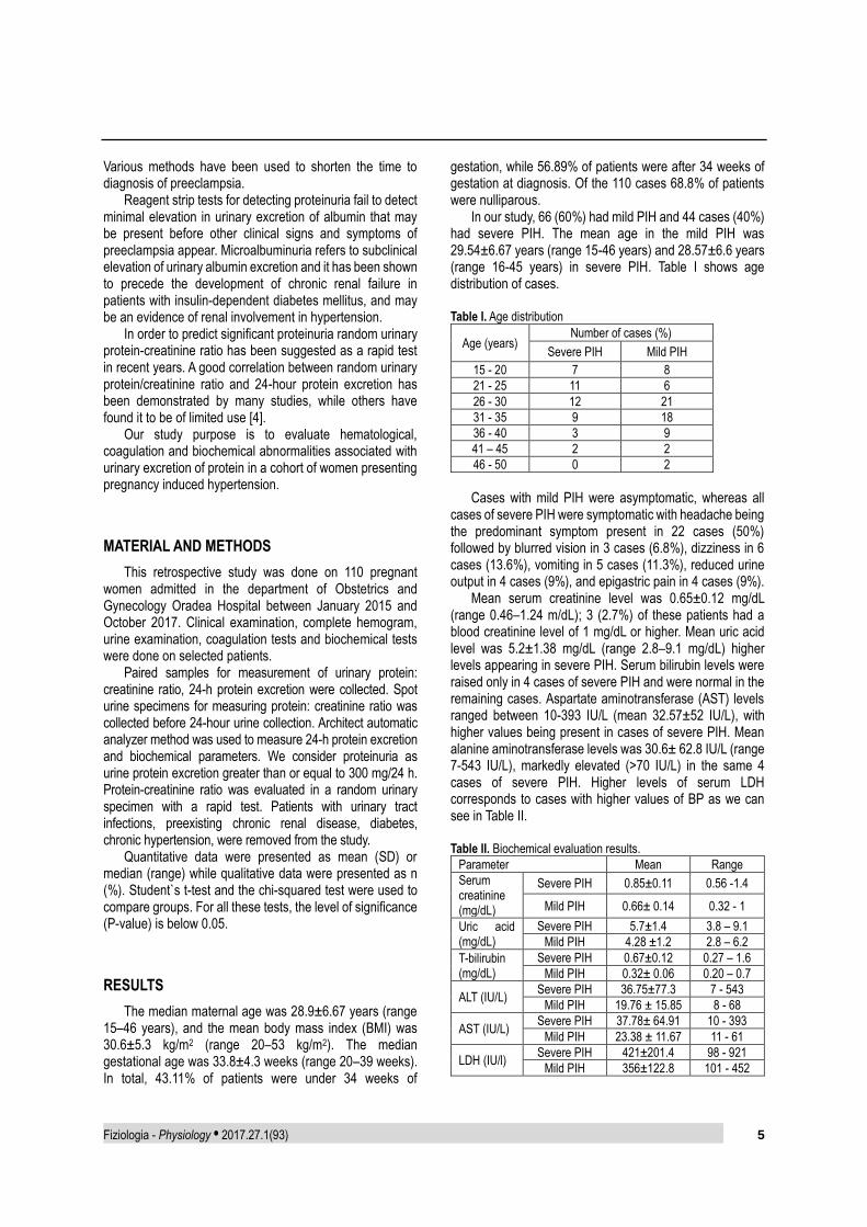
In our study, 66 (60%) had mild PIH and 44 cases (40%) had severe PIH. The mean age in the mild PIH was 29.54±6.67 years (range 15-46 years) and 28.57±6.6 years (range 16-45 years) in severe PIH. Table I shows age distribution of cases. Table I. Age distribution
Age (years) Number of cases (%)
Severe PIH Mild PIH
15 - 20 7 8
21 - 25 11 6
26 - 30 12 21
31 - 35 9 18
36 - 40 3 9
41 – 45 2 2
46 - 50 0 2
Cases with mild PIH were asymptomatic, whereas all
cases of severe PIH were symptomatic with headache being the predominant symptom present in 22 cases (50%) followed by blurred vision in 3 cases (6.8%), dizziness in 6 cases (13.6%), vomiting in 5 cases (11.3%), reduced urine output in 4 cases (9%), and epigastric pain in 4 cases (9%).
Mean serum creatinine level was 0.65±0.12 mg/dL (range 0.46–1.24 m/dL); 3 (2.7%) of these patients had a blood creatinine level of 1 mg/dL or higher. Mean uric acid level was 5.2±1.38 mg/dL (range 2.8–9.1 mg/dL) higher levels appearing in severe PIH. Serum bilirubin levels were raised only in 4 cases of severe PIH and were normal in the remaining cases. Aspartate aminotransferase (AST) levels ranged between 10-393 IU/L (mean 32.57±52 IU/L), with higher values being present in cases of severe PIH. Mean alanine aminotransferase levels was 30.6± 62.8 IU/L (range 7-543 IU/L), markedly elevated (>70 IU/L) in the same 4 cases of severe PIH. Higher levels of serum LDH corresponds to cases with higher values of BP as we can see in Table II. Table II. Biochemical evaluation results.
Parameter Mean Range
Serum creatinine (mg/dL)
Severe PIH 0.85±0.11 0.56 -1.4
Mild PIH 0.66± 0.14 0.32 - 1
Uric acid (mg/dL)
Severe PIH 5.7±1.4 3.8 – 9.1
Mild PIH 4.28 ±1.2 2.8 – 6.2
T-bilirubin (mg/dL)
Severe PIH 0.67±0.12 0.27 – 1.6
Mild PIH 0.32± 0.06 0.20 – 0.7
ALT (IU/L) Severe PIH 36.75±77.3 7 - 543
Mild PIH 19.76 ± 15.85 8 - 68
AST (IU/L) Severe PIH 37.78± 64.91 10 - 393
Mild PIH 23.38 ± 11.67 11 - 61
LDH (IU/l) Severe PIH 421±201.4 98 - 921
Mild PIH 356±122.8 101 - 452
6 Fiziologia - Physiology • 2017.27.1(93)
Hemoglobin levels of all patients varied from 7.4 to 14.35 g%, the mean value being 11.57 g%. The mean corpuscular volume (MCV) and mean corpuscular hemoglobin (MCH) in mild PIH and in severe PIH had close average values. Platelet count was between 61.080 – 466.000/mm3 in cases of severe PIH and 123.000 – 544.000/mm3 in mild PIH. The other hematological parameters did not differ significantly from those expressed in normal pregnancy that is why they were not statistically processed. Table III. Hematological parameters
Parameter Mean Range
HGB (g%) Severe PIH 11.68 ± 1.6 7.4 – 12.35
Mild PIH 11.43±1.53 8.57 – 14.11
MCV Severe PIH 83.06±6.25 62.3 - 94
Mild PIH 83.38±6.01 70 - 99
MCH Severe PIH 27.25±2.75 18 – 33.5
Mild PIH 27.5±2.48 22 - 32
PLT Severe PIH 249±80.34 61.08 - 466
Mild PIH 269±105.65 152 - 544
Concerning the coagulation tests, fibrinogen was increased in all cases, mean value 676.29±200.67mg/dL. APTT was normal in all cases of mild PIH and most cases of severe PIH but prolonged (>29 s) in 6 cases of the last one. The PT was significantly prolonged only in 4 cases of severe PIH. Table IV. Coagulation tests results
Parameter Mean Range
Fibrinogen (mg/dl)
Severe PIH
662.79 ±197.19
198 - 1388
Mild PIH 700.2±208.3 256 - 1228
APTT (sec)
Severe PIH
24.7±2.75 18.9 – 32.5
Mild PIH 25.78±3.2 18.1 – 32.2
PT (sec)
Severe PIH
8.79±1.13 5.4 – 13.5
Mild PIH 9.16±0.75 7.8 – 11.3
The mean protein level was 3.94 g (range 0.1–13 g) in 24-hour protein excretion test for 110 patients. Values were higher in the severe PIH group. The mean P/C ratio was 3.16 mg/g (range 0.12–69.6 mg/g). Urine examination revealed 1+ and 2+ proteinuria in 48 cases (72.72%) of mild PIH, while majority (41 cases, 93.18%) of those with severe PIH had 2+ and 3+ proteinuria. Table V. Urinary results
Parameter
Urinary protein (mg/24h)
Severe PIH 2866.28±3328.81
Mild PIH 886.67±525.6
Spot urine protein (mg/dL)
Severe PIH 314.68± 257.73
Mild PIH 145.90±107.79
Spot urine creatinine (mg/dL)
Severe PIH 92.03± 68.87
Mild PIH 85.09±49.65
Protein/creatinine ratio (mg/mg)
Severe PIH 3.17±0.79
Mild PIH 2.92± 0.48
DISCUSSION
In the present study, the mean age in the mild PIH group was 29.49 ± 6.66 years and, in the severe PIH group it was 28±6.67 years, being in concordance with the observation made by O’Brien et al. that PIH is mainly a disease of young nulliparous [5]. Headache was the predominant symptom seen in 50% of severe PIH patients followed by dizziness in 13.6% of patients, the results being similar with the one reported in other studies.
Abnormal serum bilirubin levels were detected as result of liver damage, including because of pre-eclampsia, its rise signifying the severity of the condition. On the other hand, bilirubin is described as an anti-oxidant and levels in the upper end of the normal range are associated with reduced cardiovascular and respiratory morbidity [6]. It has been suggested that a higher oxidative stress response means a more severe form of pre-eclampsia, therefore consumption of more anti-oxidants e.g. bilirubin. Many studies reported mean bilirubin concentration in severe PIH to be significantly higher than in mild PIH. In our study, there was no significant difference in mean bilirubin concentration between the mild and severe PIH groups; levels have been raised only in 4 cases of severe PIH.
Serum LDH levels has significant associations with various disorders, therefore total serum LDH is a sensitive but a nonspecific test. In preeclampsia also LDH may be elevated and can indicate the prognosis for both mother and fetus [7]. Because levels of LDH < 600 IU/L are common in normal pregnancy only levels > 800 IU/L were reported to be associated with complicated pre-eclampsia [8]. In the present study the LDH levels raised proportionally with the increase of the values of DP and BP, but only 4 cases showed values over 800 IU/l, in the severe PIH group.
The mean AST, ALT values were higher than normal (>70 IU/L) in severe PIH group, four cases presented values greater than 100 IU/L. Liver enzymes may contribute to the diagnosis of HELLP syndrome in addition with the other biochemical and hematological parameters.
During early pregnancy serum uric acid level decreases, related to the effects of estrogen and the increase in renal blood flow. Uric acid levels then increase during the third trimester, reaching levels of 4–5 mg/dL by term. It is known that subjects who develop preeclampsia show slightly higher serum uric acid levels during the first trimester in association with a relative reduction in urinary urate excretion and the degree of elevation correlates with the severity of the maternal and fetal complications [9]. In our study, 30 patients over 20 weeks of pregnancy had elevated uric acid levels (>8 mg/dL), 21 belonging to severe PIH group, with values between 6 and 9.1 mg/dL. The remaining cases with elevated uric acid, between 6-8.2 mg/dL belonged to the mild PIH group.
Women with pre-eclampsia develop a variety of hematologic dysfunctions such as platelet abnormalities, red cells abnormalities with impact on the maternal and neonatal
Fiziologia - Physiology • 2017.27.1(93) 7
morbidity and mortality. Patients with severe pre-eclampsia have a microangiopathic hemolytic anemia. The mean values of hemoglobin, MCV and MCH were close in the two PIH groups. Patients with anemia were included in the iron deficiency cause of microscopic hypochromic anemia (25 from 110 cases). Hemolytic peripheral blood picture was present in only four cases of severe PIH, confirmed by findings on peripheral smear (burr cells, schistocytes, polychromasia). All these four cases had thrombocytopenia and elevated liver enzymes.
The platelet count decreases in normal pregnancy possibly due to haemodilution, with a maximal decrease in the third trimester. On the other hand, thrombocytopenia is a common hemostatic abnormality of preeclampsia, present in approximately 50% of patients according to a recent study done by Donimath et al. [10] In our study, 10% patients had thrombocytopenia and they were part of the severe PIH group. There is a significant relationship between thrombocytopenia and the severity of the PIH.
Pregnancy is associated with changes in hemostasis, an increase in the level of clotting factors, a decrease in the quantity of natural anticoagulants and a reduction in fibrinolytic activity. Pregnancy leads to a state of hypercoagulability, likely due to hormonal changes and increases the risk of thromboembolism. Coagulation profile is an important parameter for detecting effects of PIH or pre-eclampsia, patients could have a significant abnormality in platelet function as well as number. Coagulation studies were done in all the patients included in our study. The mean PT was significantly prolonged in cases with severe PIH, according with similar observation made by others [11] The mean APTT in severe PIH was significantly prolonged in the study done by Thomas et al. and Jambhulkar et al. [11,12]. There were no significant differences between the mean APTT of mild and severe PIH patients in our study.
Proteinuria is a diagnostic test of pre-eclampsia. In our study, proteinuria was present in all the cases (100%). Using 24-hour protein collection, Jambhulkar et al. observed proteinuria in only 68% cases of mild PIH and 92% cases of severe PIH [12]. The P/C ratio in spot urine was introduced as an alternative test. Many studies reported potential errors in determining protein in a spot urine sample due to the daily variation of protein elimination. Evaluation of spot urine P/C ratio in pregnant women with suspected preeclampsia has been found to be closely correlated with the 24-hour urine protein measurement, but there is no reliable evidence about the optimal cut-off value for defining it. The most recent study stated that the optimum threshold for P/C ratio to determine proteinuria is between 0.30 and 0.35, with determining sensitivity and specificity values above 75%; when the sensitivity and specificity above 80% was accepted, there was no cut-off found [13]. In our study, urine parameters examined have shown higher values in the severe PIH group.
CONCLUSION
Younger patients with lower parity grade had significantly increased systolic and diastolic blood pressure values and elevations of values of liver enzymes, uric acid, urine protein, and LDH levels. Abnormalities (qualitative and quantitative abnormalities) of red blood cells and platelets are frequently seen in PIH. Coagulation tests can be changed because of thrombocytopenia, as increased platelet consumption is an early feature of this disorder. We conclude that spot urinary P/C ratio in hospitalized women with suspected preeclampsia can be used as a screening test and a good predictor for significant proteinuria but cannot be used as diagnostic tests to replace 24-h urine protein measurements.
REFERENCES
1. Palma Gamiz JL. Arterial hypertension and pregnancy: Diagnostic criteria and therapeutic approach. Rev Esp Cardiol 514: 50-58, 1998.
2. Padden MO. HELLP syndrome: Recognition and perinatal management, Am Fam Physician 1999; 60:829-36, 839.
3. Tranquilli A, Dekker G, Magee L, et al. The classification, diagnosis and management of the hypertensive disorders of pregnancy: a revised statement from the ISSHP. Hypertens Pregnancy. 2014; 4:97-104.
4. Stout MJ, Scifres CM, Stamilio DM. Diagnostic utility of urine protein-to-creatinine ratio for identifying proteinuria in pregnancy, J Matern Fetal Neonatal Med. 2013 Jan; 26(1): 66-70.
5. O’Brien WF, Saba HI, Knuppel RA, Scerbo JC, Cohen GR. Alterations in platelet concentration and aggregation in normal pregnancy and preeclampsia. Am J Obstet Gynecol 1986; 155:486-90.
6. Wang L, Bautista LE. Serum bilirubin and the risk of hypertension. Int J Epidemiol 2015; 44(1).
7. Jaiswar SP, AmritG, Rekha S, Natu SN, Mohan S. Lactic Dehydrogenase: A Biochemical Marker for Preeclampsia–Eclampsia. The Journal of Obstetrics and Gynecology of India, 2011; 61(6):645-648.
8. Vinitha Padmini Mary, Chellatamizh M, Padmanaban S. Role of serum LDH in preeclampsia as a prognostic factor – a cross sectional case control study in tertiary care hospital. Int J Reprod Contracept Obstet Gynecol. 2017; 6(2):595-598.
9. Johnson RJ, Kanbay M, Kang DH, Sánchez Lozada LG, Feig D. Uric acid: A Clinically Useful Marker to Distinguish Preeclampsia from Gestational Hypertension. Hypertension 2011; 58(4): 548-549.
10. Donimath KV, Sambrani AM, Rathod PM. A study on association of thrombocytopenia with pregnancy induced hypertension. Int J Reprod Contracept Obstet Gynecol. 2016; 5(3):808-812.
11. Thomas A, Hanagavadi S, Kadam S, Shashikala P, Chandrashekhar HR. Coagulation profile in PIH. Indian J Hemat Blood Trans 1998; 16:7-11.
12. Jambhulkar S, Shrivastava R, Shrikhande A, Deshmukh K. Coagulation profile in pregnancy induced hypertension, Indian Journal of Hematology and Blood Transfusion 2001; 19(1):3-5.
8 Fiziologia - Physiology • 2017.27.1(93)
13. Demirci O, Kumru P, Arınkan A, et al. Spot Protein/Creatinine Ratio in Preeclampsia as an
Alternative for 24-Hour Urine Protein. Balkan Med J 2015; 32:51-5.
EVALUAREA CLINICO-PATOLOGICĂ A HIPERTENSIUNII INDUSE DE SARCINĂ
REZUMAT Introducere. Deoarece manifestările clinice ale preeclampsiei pot fi heterogene, testele de laborator sunt foarte utile în diagnosticul precoce al preeclampsiei la pacientele cu hipertensiune indusă de sarcină. Metode. În acest studiu au fost incluse 110 paciente cu diagnostic clinic de hipertensiune arterială indusă de sarcină (PIH). S-au efectuat următoarele analize la toate pacientele: hemoleucogramă, examen sumar de urină, teste biochimice (ALT, AST, LDH, bilitubina totală, acid uric) și teste de coagulare. Testele urinare au inclus parametrii uzuali și determinarea proteinuriei (în urina colectată 24h). În plus, a fost determinat și raportul proteine/creatinină (P/C) în probele de urină cu ajutorul unui test rapid. Rezultate. În acest studiu, 66 de paciente au prezentat PIH ușoară și 44 de paciente au avut PIH severă. Cefaleea a fost simptomul predominant, urmat de amețeli. Modificările patologice ale parametrilor hematologici, biochimici și de coagulare au fost observate mai ales la pacientele cu PIH severă. Concluzii. Pacientele semnificativ mai tinere, cu paritate scăzută au prezentat valori semnificativ crescute ale tensiunii sistolice și diastolice și modificări ale valorilor enzimelor hepatice, acidului uric, proteinelor urinare și LDH. Anomalii (calitative și cantitative) ale eritrocitelor și trombocitelor sunt întâlnite frecvent în PIH. Testul rapid de determinare a raportului P/C din urină la pacientele cu hipertensiune indusă de sarcină poate fi folosit ca test de screening în predicția proteinuriei semnificative și nu poate fi utilizat ca test diagnostic în locul evaluării proteinuriei/24 h. Cuvinte cheie: sarcină, hipertensiune, preeclampsie, diagnostic.
Fiziologia - Physiology • 2017.27.1(93) 9
HYPERURICEMIA – THE NEW CARDIOVASCULAR
RISK FACTOR - CONSEQUENCES AND EFFECTS
MĂDĂLINA IOANA MOISI1
, COSMIN MIHAI VESA1
, ADRIANA IOANA ARDELEAN2
, DANA
ZAHA2
, CISMAŞ PETRIŞOR PRUTEANU2
, IOAN MAGYAR2
, MIRCEA IOACHIM POPESCU2
1Emergency County Hospital Oradea 2Faculty of Medicine and Pharmacy, University of Oradea
ABSTRACT Introduction: The concept of correlation between serum uric acid high levels and cardiovascular disease is strongly debated lately and remains controversial. Different authors believe that uricemia is a risk factor, prediction factor or even causal factor. Interpretation of data is difficult because of strong interrelations between serum uric acid and all CV risk factors, especially when metabolic syndrome is associated. Material and Methods: We gathered the documents of patients dispensed from the cardiology department of the County Emergency Clinical Hospital from Oradea, from 2016, and we formed a study lot consisting of 25 patients. The study lot was divided in two gruops, so the subjects with normal serum level of uric acid represented the control group (C.G), and the patients with hyperuricemia were included in the refference group (R.G). Clinical examination, laboratory tests , electocardiography and 2D echocardiography were also performed. Results: The C.G. developped especially grade I and II hypertension, stable angina pectoris and heart failure NYHA I and II. This fact reflects the correlation of hyperuricemia with high blood pressure ( grade III) in 33.33% of the R.G. , unstable angina pectoris in 26,66% of the R.G., and heart failure NYHA III and IV was discovered in 40% and 20 % of the subjects from the R.G. Acute myocardial infarction was detected only in the R.G. The explanation reflects that uric acid under ischemic conditios will generate oxidative stress reactive species (peroxynitrite) promoting endothelial disfunction, inflammation, oxidized LDL synthese which will be storaged in the foamy cells from the initmal layers of the vessels, so the atheroma plaque will be intiated. Conclusions: Even if the quality of risk factor isn’t well defined, it is mandatory that a regular laboratory examination include the uric acid serum levels. In the global assessment of cardiovascular risk factors , uric acid determination may be helpful, providing dates for estabilishing a long therm prognosis and future therapeutic intervention. In patients with cardiovascular pathology who have a high CV risk, the detection and management of hyperuricemia has become essential. European guidelines regarding the heart failure and coronary artery disease recommends treatment with allopurinol in patients with hyperuricemia. Keywords: Hiperuricemie, endothelial disfunction, reactive species of oxygen, inflammation
INTRODUCTION
Important epidemiological studies have demonstrated the association of serum uric acid high levels with hypertension, cerebrovascular disease, coronary artery disease, preeclampsia and renal disease [1].
Analyzing the data of numerous prospective studies investigating the association of serum uric levels in different subjects, most authors concluded that in the general population at low CV risk, if the traditional risk factors are eliminated, uric acid becomes a poor morbidity predictor. In hypertensive and especially in high and high risk CV categories (in diabetics, in the case of a history of stroke, in those with coronary angiographic disease or in patients with heart failure), uric acid has been shown to be a major CV risk predictor [2].
The release of adenosine is increased under hypoxia conditions and its vasodilating action will restore local blood flow thus preventing tissue ischemia. The final adenosine catabolism product is uric acid, thus hyperuricemia became an important marker for detecting subclinical ischemia.
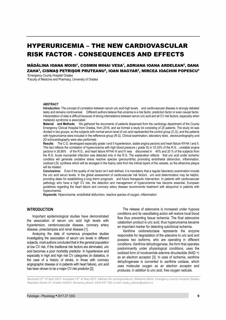
Xanthine oxidoreductase represents the enzyme responsible for degradation of the adeosine to uric acid and possess two isoforms, who are operating in different conditions. Xanthine dehydrogenase, the form that operates predominantly under physiological conditions, uses the oxidized form of nicotinamide adenine dinucleotide (NAD +) as an electron acceptor [3]. In case of ischemia, xanthine dehydrogenase is converted to xanthine oxidase, which uses molecular oxygen as an electron acceptor and produces, in addition to uric acid, free oxygen radicals.
Received 22nd of April 2017. Accepted 13th of May 2017. Address for correspondence: Madalina Moisi, Emergency County Hospital Oradea, Republicii Street 37, Oradea 410167, Romania; phone: 0259 437 750; e-mail: [email protected]
The high uric acid will promote oxidized LDL particles
synthesis through these oxygen stress reactive species (peroxynitrite) [4]. These molecules will be stored in the foamy cells in the intimal layers of the vessels, consisting the first step in developing the atheroma plaque.
Another source of production for foam cells is smooth muscle cells from medium layer of the vessels. Unlike macrophages, smooth muscle cells have a higher expression of LDL receptors on their surface. Both cell types have frenzied mechanisms that prevent excessive incorporation of cholesterol, but accumulation of cholesterol is more pronounced in atherosclerotic areas. It is assumed that apo-E with increased affinity for LDL favors macrophage metamorphosis [5].
Modified LDL stored in the intimal layers inhibits NO production and stimulates ET1 synthesis, inhibits the release of prostacyclin favoring thromboxane production, augmenting vasoconstriction and platelets aggregation.
Another consequence of high uric acid and oxidative reactive species production is the release of NF-IB (nuclear transcription factor) which is responsible for inflammation through increased cytokines synthesis [6].
The family of adhesion molecules can be divided into three categories: selectins (L-selectin, E-selectin, P-selectin), integrins and immunoglobulins (VCAM, ICAM, PECAM-1) [4]. Adhesion of endothelial monocytes, platelets and neutrophils leads to so-called endothelial activation with various consequences: synthesis of pro-inflammatory cytokines by activated macrophages, T lymphocytes, endothelial cells, smooth muscle cells; surface receptor production, enzymes, growth factors. Inflammatory cytokines are interleukins, TNF, MCP-1, MCP-4, COX-2 [4]. Inflammation is responsible for cellular apoptosis and uric acid production.
MATERIAL AND METHODS
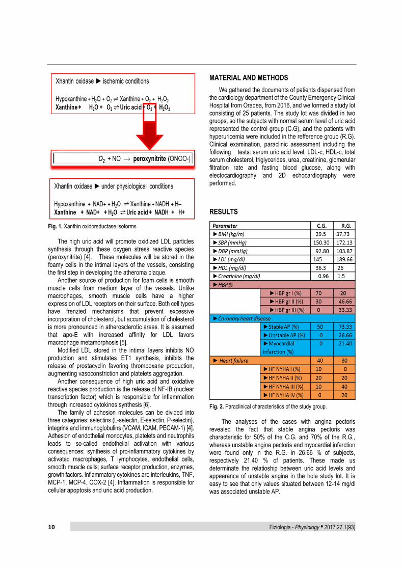
We gathered the documents of patients dispensed from the cardiology department of the County Emergency Clinical Hospital from Oradea, from 2016, and we formed a study lot consisting of 25 patients. The study lot was divided in two gruops, so the subjects with normal serum level of uric acid represented the control group (C.G), and the patients with hyperuricemia were included in the refference group (R.G). Clinical examination, paraclinic assessment including the following tests: serum uric acid level, LDL-c, HDL-c, total serum cholesterol, triglycerides, urea, creatinine, glomerular filtration rate and fasting blood glucose, along with electocardiography and 2D echocardiography were performed.
RESULTS
Fig. 2. Paraclinical characteristics of the study group.
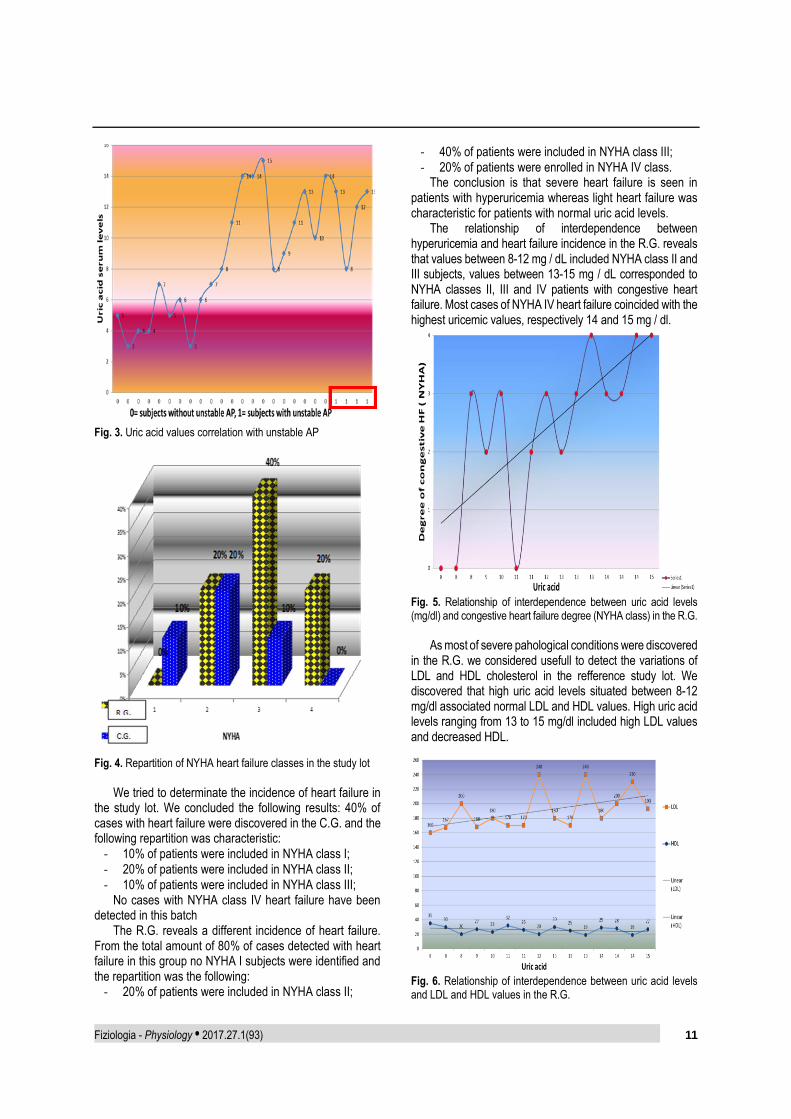
The analyses of the cases with angina pectoris
revealed the fact that stable angina pectoris was characteristic for 50% of the C.G. and 70% of the R.G., whereas unstable angina pectoris and myocardial infarction were found only in the R.G. in 26.66 % of subjects, respectively 21.40 % of patients. These made us determinate the relatioship between uric acid levels and appearance of unstable angina in the hole study lot. It is easy to see that only values situated between 12-14 mg/dl was associated unstable AP.
Fiziologia - Physiology • 2017.27.1(93) 11
Fig. 3. Uric acid values correlation with unstable AP
Fig. 4. Repartition of NYHA heart failure classes in the study lot
We tried to determinate the incidence of heart failure in
the study lot. We concluded the following results: 40% of cases with heart failure were discovered in the C.G. and the following repartition was characteristic:
‐ 10% of patients were included in NYHA class I; ‐ 20% of patients were included in NYHA class II; ‐ 10% of patients were included in NYHA class III;
No cases with NYHA class IV heart failure have been detected in this batch
The R.G. reveals a different incidence of heart failure. From the total amount of 80% of cases detected with heart failure in this group no NYHA I subjects were identified and the repartition was the following:
‐ 20% of patients were included in NYHA class II;
‐ 40% of patients were included in NYHA class III; ‐ 20% of patients were enrolled in NYHA IV class.
The conclusion is that severe heart failure is seen in patients with hyperuricemia whereas light heart failure was characteristic for patients with normal uric acid levels.
The relationship of interdependence between hyperuricemia and heart failure incidence in the R.G. reveals that values between 8-12 mg / dL included NYHA class II and III subjects, values between 13-15 mg / dL corresponded to NYHA classes II, III and IV patients with congestive heart failure. Most cases of NYHA IV heart failure coincided with the highest uricemic values, respectively 14 and 15 mg / dl.
Fig. 5. Relationship of interdependence between uric acid levels (mg/dl) and congestive heart failure degree (NYHA class) in the R.G.
As most of severe pahological conditions were discovered
in the R.G. we considered usefull to detect the variations of LDL and HDL cholesterol in the refference study lot. We discovered that high uric acid levels situated between 8-12 mg/dl associated normal LDL and HDL values. High uric acid levels ranging from 13 to 15 mg/dl included high LDL values and decreased HDL.
Fig. 6. Relationship of interdependence between uric acid levels and LDL and HDL values in the R.G.
R.G.
12 Fiziologia - Physiology • 2017.27.1(93)
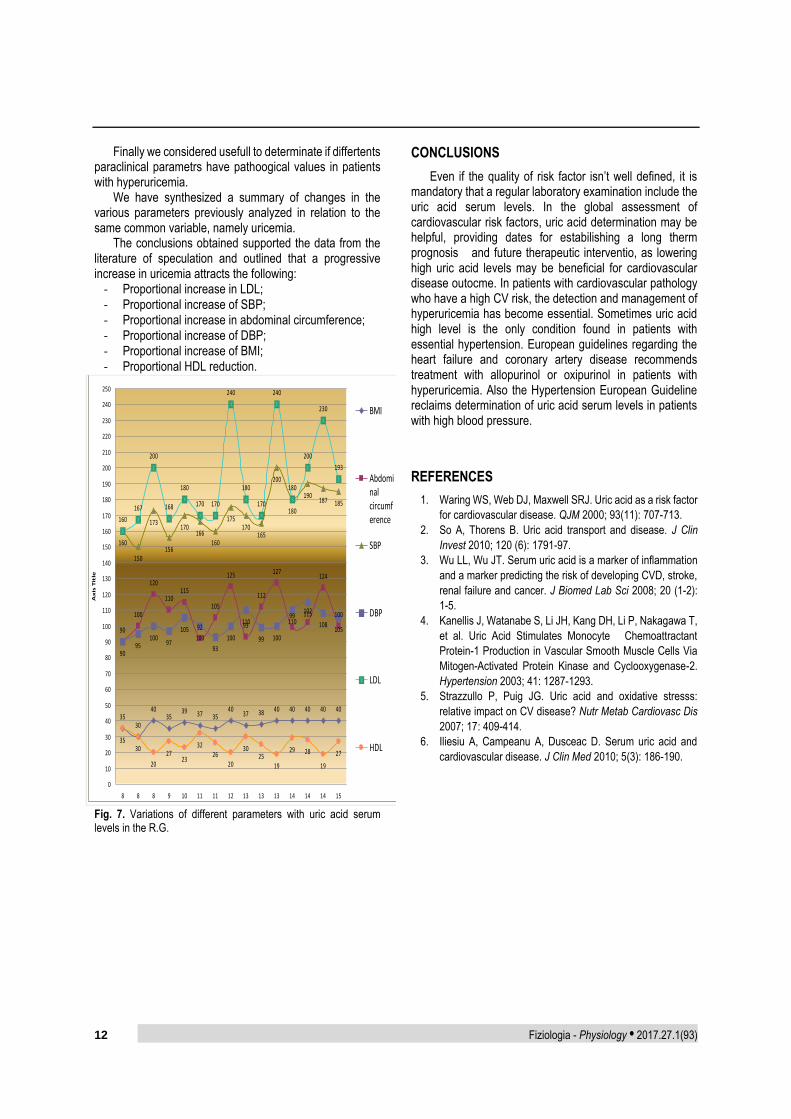
Finally we considered usefull to determinate if differtents paraclinical parametrs have pathoogical values in patients with hyperuricemia.
We have synthesized a summary of changes in the various parameters previously analyzed in relation to the same common variable, namely uricemia.
The conclusions obtained supported the data from the literature of speculation and outlined that a progressive increase in uricemia attracts the following:
‐ Proportional increase in LDL; ‐ Proportional increase of SBP; ‐ Proportional increase in abdominal circumference; ‐ Proportional increase of DBP; ‐ Proportional increase of BMI; ‐ Proportional HDL reduction.
Fig. 7. Variations of different parameters with uric acid serum levels in the R.G.
CONCLUSIONS
Even if the quality of risk factor isn’t well defined, it is mandatory that a regular laboratory examination include the uric acid serum levels. In the global assessment of cardiovascular risk factors, uric acid determination may be helpful, providing dates for estabilishing a long therm prognosis and future therapeutic interventio, as lowering high uric acid levels may be beneficial for cardiovascular disease outocme. In patients with cardiovascular pathology who have a high CV risk, the detection and management of hyperuricemia has become essential. Sometimes uric acid high level is the only condition found in patients with essential hypertension. European guidelines regarding the heart failure and coronary artery disease recommends treatment with allopurinol or oxipurinol in patients with hyperuricemia. Also the Hypertension European Guideline reclaims determination of uric acid serum levels in patients with high blood pressure.
REFERENCES
1. Waring WS, Web DJ, Maxwell SRJ. Uric acid as a risk factor
for cardiovascular disease. QJM 2000; 93(11): 707-713.
2. So A, Thorens B. Uric acid transport and disease. J Clin
Invest 2010; 120 (6): 1791-97.
3. Wu LL, Wu JT. Serum uric acid is a marker of inflammation
and a marker predicting the risk of developing CVD, stroke,
4. Kanellis J, Watanabe S, Li JH, Kang DH, Li P, Nakagawa T,
et al. Uric Acid Stimulates Monocyte Chemoattractant
Protein-1 Production in Vascular Smooth Muscle Cells Via
Mitogen-Activated Protein Kinase and Cyclooxygenase-2.
Hypertension 2003; 41: 1287-1293.
5. Strazzullo P, Puig JG. Uric acid and oxidative stresss:
relative impact on CV disease? Nutr Metab Cardiovasc Dis
2007; 17: 409-414.
6. Iliesiu A, Campeanu A, Dusceac D. Serum uric acid and
cardiovascular disease. J Clin Med 2010; 5(3): 186-190.
3530
4035
39 37 3540
37 38 40 40 40 40 40
90
100
120
110115
92
105
125
93
112
127
99102
124
100
160
150
173
156
170166
160
175170
165
200
180
190187 185
9095
10097
105100
93
100
110
99 100
110115
108105
160
167
200
168
180
170 170
240
180
170
240
180
200
230
193
3530
20
2723
3226
20
3025
19
29 28
19
27
0
10
20
30
40
50
60
70
80
90
100
110
120
130
140
150
160
170
180
190
200
210
220
230
240
250
8 8 8 9 10 11 11 12 13 13 13 14 14 14 15
Ax
is T
itle
BMI
Abdominalcircumference
SBP
DBP
LDL
HDL
Fiziologia - Physiology • 2017.27.1(93) 13
HIPERURICEMIA ASIMPTOMATICA - NOUL FACTOR DE RISC
CARDIOVASCULAR? - CONSECINȚE ȘI EFECTE
REZUMAT Introducere: Conceptul de corelație între valorile ridicate ale acidului uric seric și bolile cardiovasculare este dezbătut în ultimul timp și rămâne controversat. Diferiți autori confirmă faptul că hiperuricemia constituie un factor de risc, un factor de predicție sau chiar un factor cauzal. Interpretarea datelor este dificilă datorită interacțiunilor puternice dintre acidul uric seric și toți factorii de risc CV, mai ales când se asociază si sindromul metabolic. Material și Metode: Am selectat documentele pacienților externaţi din departamentul de cardiologie al Spitalului Clinic Județean de Urgență din Oradea, din anul 2016, și am format un lot de studiu format din 25 de pacienți. Lotul de studiu a fost împărțit în două grupe, subiecții cu un nivel normal al acidului uric în serul seric au reprezentat grupul de control (C.G), iar pacienții cu hiperuricemie au fost incluși în grupul de referință (R.G). Au fost de asemenea efectuate examinări clinice, rezultate de laborator, electrocardiografie și ecocardiografie 2D. Rezultate: C.G. au dezvoltat în special hipertensiune arterială grad I şi II, angină pectorală stabilă și insuficiență cardiacă NYHA I și II. Acest fapt reflectă corelarea hiperuricemiei cu tensiunea arterială ridicată (gradul III) în 33,33% din cazuri din R.G., angina pectorală instabilă în 26,66% din cazuri, de la R.G., și insuficiența cardiacă NYHA III și IV la 40% , respectiv 20% din pacienții din RG. Infarctul miocardic acut a fost detectat detectat numai în RG Explicația reflectă faptul că acidul uric ,în conditii ischemice, va genera specii reactive de stres oxidativ (peroxinitrit) care promovează disfuncția endotelială, inflamația, sinteza particulelor LDL oxidate care vor fi depozitate la nivelul celulelor spumoase din itraturile intimale ale vaselor, formându-se astfel placa de aterom. Concluzii: Chiar dacă rolul de factor de risc nu este bine definit, este obligatoriu ca un examen de laborator să includă și detectarea nivelurile serice ale acidului uric. În evaluarea globală a factorilor de risc cardiovasculari, determinarea acidului uric poate fi utilă, oferind date pentru stabilirea unui prognostic şi viitoarele ţinte terapeutice. În cazul pacienților cu patologie cardiovasculară și risc crescut CV, detectarea și gestionarea hiperuricemiei au devenit esențiale. Ghidurile europene de insuficiența cardiacă și boala coronariană recomandă tratamentul cu alopurinol la pacienții cu hiperuricemie. Cuvinte cheie: Hiperuricemie, disfuncție endoteliala, specii reactive ale oxigenului, inflamație
14 Fiziologia - Physiology • 2017.27.1(93)
IMPLICATION OF DISTRESS AND EMOTIONAL
INTELLIGENCE IN FORENSIC PRACTICE
ADRIANA COJOCARU, LUMINIȚA AGEU
Center of Mental Health for Children and Adolescents, Timisoara, Romania
ABSTRACT The present paper aims to investigate the relationship between empathy and distress, as well as their implications in the activity of the forensic practitioners. The study was conducted on a group of 30 doctors. Emotional distress was measured using the Profile of Affective Distress (PDA), and the Emotional Intelligence Scale (SEI) was used to evaluate the level of empathy and the way subjects manage emotions. The results showed that both women and men are equally emphatic, with no differences when it comes to regulating the emotions of others and emotions evaluation. Keywords: emotional intelligence, forensic, distress.
INTRODUCTION
The medical-forensic clinic and pathology are the two cutting-edge medical disciplines where doctors often receive information that is agonizing and traumatically from a psychological point of view, first-hand information, in the form of historical results, of examination and investigation. They do not receive the information in an ad-hoc manner (as is the case with the emergency department physicians), but in a daily continuous way, throughout their active career [1, 2].
Their short and long term implications on the physician's psychological health as well as the defence mechanisms in the medic's way of thinking and the implications on their personality and behaviour have been very poorly researched [3-5].
If the doctor does not use his wisdom and the experience obtained over the years in order to cope with the distress, he may be overwhelmed by the situation and his opinion may become subjective, biased. The emotional lability of a doctor may lead him on some unprofessional ways.
MATERIAL AND METHODS
A group of 30 people aged between 36 and 60, having a work experience between 10 and 40 years, forensic experts were asked to complete two questionnaires regarding emotional distress and emotional intelligence. The questionnaires used were: Profile of Affective Distress (PDA) and Emotional Intelligence (SEI), each scale containing several other subscales. The affective distress scale is a tool developed for the purpose of assessing the subjective dimension of negative functional emotions, dysfunctional
negative emotions, and positive emotions. The Emotional intelligence scale comprises 4 subscales: Emotion Assessment; adjusting one's personal emotions; adjusting the emotions of others and the utilisation of emotions [6].
The average age of the subject group is 48 years, of which 12 are female, with the remaining 18 subjects being male.
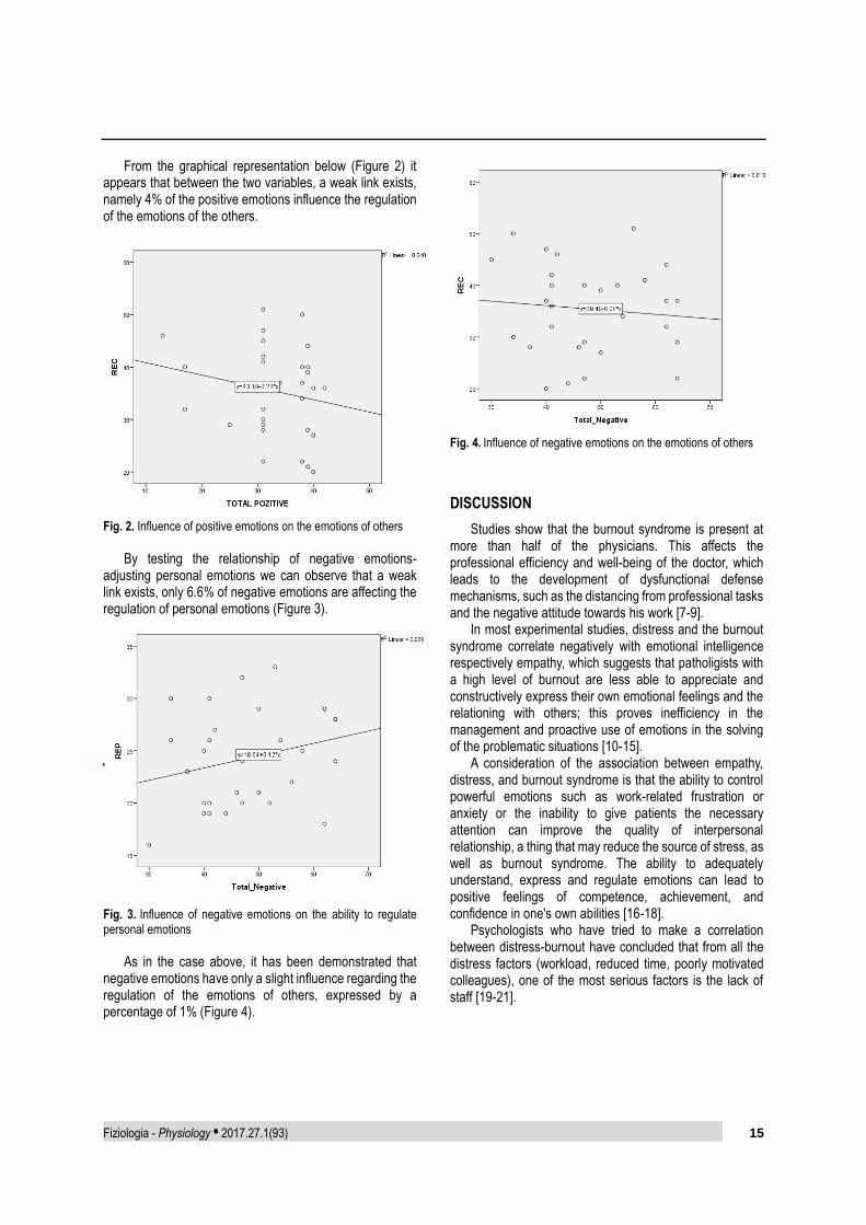
RESULTS
Figure 1 shows that there is a weak link between the two variables; only 7.7 percent of positive emotions have an influence on the regulation of personal emotions.
Fig. 1. Influence of positive emotions on the ability to regulate personal emotions
Received 10th of March 2017. Accepted 12th of April 2017. Address for correspondence: Luminița Ageu, PhD, Center of Mental Health for Children and Adolescents, Corbului Street no. 7, Timisoara, Romania; phone: +0723056089; e-mail: [email protected]
From the graphical representation below (Figure 2) it appears that between the two variables, a weak link exists, namely 4% of the positive emotions influence the regulation of the emotions of the others.
Fig. 2. Influence of positive emotions on the emotions of others
By testing the relationship of negative emotions-
adjusting personal emotions we can observe that a weak link exists, only 6.6% of negative emotions are affecting the regulation of personal emotions (Figure 3).
Fig. 3. Influence of negative emotions on the ability to regulate personal emotions
As in the case above, it has been demonstrated that
negative emotions have only a slight influence regarding the regulation of the emotions of others, expressed by a percentage of 1% (Figure 4).
Fig. 4. Influence of negative emotions on the emotions of others
DISCUSSION
Studies show that the burnout syndrome is present at more than half of the physicians. This affects the professional efficiency and well-being of the doctor, which leads to the development of dysfunctional defense mechanisms, such as the distancing from professional tasks and the negative attitude towards his work [7-9].
In most experimental studies, distress and the burnout syndrome correlate negatively with emotional intelligence respectively empathy, which suggests that patholigists with a high level of burnout are less able to appreciate and constructively express their own emotional feelings and the relationing with others; this proves inefficiency in the management and proactive use of emotions in the solving of the problematic situations [10-15].
A consideration of the association between empathy, distress, and burnout syndrome is that the ability to control powerful emotions such as work-related frustration or anxiety or the inability to give patients the necessary attention can improve the quality of interpersonal relationship, a thing that may reduce the source of stress, as well as burnout syndrome. The ability to adequately understand, express and regulate emotions can lead to positive feelings of competence, achievement, and confidence in one's own abilities [16-18].
Psychologists who have tried to make a correlation between distress-burnout have concluded that from all the distress factors (workload, reduced time, poorly motivated colleagues), one of the most serious factors is the lack of staff [19-21].
16 Fiziologia - Physiology • 2017.27.1(93)
CONCLUSION
Analysing the scientific literature as well as the results of the current study, it was concluded that age positively influences the emotional intelligence, respectively empathy, and adversely distress.
The current study may provide useful data for some future experimental work. A future perspective may represent the analysis of the correlations between the many variables existing in the paper.
REFERENCES
1. Diaconescu M. The Concept of Empathy in Philosophy and Psychotherapy. Babeş-Bolyai University Cluj, 2008,105-115.
2. Nilsson P. Empathy and Emotions. On The Notion of Empathy as Emotional Sharing. Ůmea Studies in Philosophy 7, Gunnar Andersson, Ingvar Johansson and Sten Lindstrom eds., Department of Philosophy and Linguistic, Umea Unversity, Sweden, 2003.
3. Nussbaum LA, Hogea LM, Andreescu NI, Grădinaru RC, Puiu M, Todica A. The Prognostic and Clinical Significance of Neuroimagistic and Neurobiological Vulnerability Markers in Correlation with the Molecular Pharmacogenetic Testing in Psychoses and Ultra High-Risk Categories. Rom J Morphol Embryol 2016, 57(3): 959-967.
4. Nussbaum L, Gradinaru R, Andreescu N, Dumitrascu V, Tudor A, Suciu L, Stefanescu R, Puiu M. The Response to Atypical Antipsychotic Drugs in Correlation with the CYP2D6 Genotype: Clinical Implications and Perspectives. Farmacia 2014, 62(6): 1191-1201.
5. Nussbaum L, Hogea LM, Călina D, Andreescu N, Grădinaru R, Ștefănescu R, Puiu M. Modern Treatment Approaches in Psychoses. Pharmacogenetic, Neuroimagistic and Clinical Implications. Farmacia 2017, 65(1), 75-81.
6. Dymond R. A scale for the measurement of empathic ability. J Consult Psychol 1949, 13:127-133.
7. Battin D. Book reviw on ”Psychoanalytic Empathy”, by Stefano Bolognini. In: JAPA 2005; 53: 1010-1014.
8. Maibom HL. Empathy: Philosophical and Psychological Perspectives, edited by Amy Coplan and Peter Goldie, 2014, 123-491.
9. Shirom A. Job-related burnout: A review, In Quick JC, Tetrick LE (eds.), Handbook of Occupational Health Psychology, American Psychological Association, Washigton, DC, 2009.
10. Timar B, Popescu S, Timar R, Baderca F, Duica B, Vlad M, Levai C, Balinisteanu B, Simu M. The usefulness of quantifying intraepidermal nerve fibers density in the diagnostic of diabetic peripheral neuropathy: a cross-sectional study. Diabetol Metab Syndr 2016, 8:31.
11. Timar B, Timar R, Gaita L, Oancea C, Levai C, Lungeanu D. The impact of diabetic neuropathy on balance and on the risk of falls in patients with type 2 diabetes mellitus: a cross-sectional study. PLoS One 2016, 11(4): e0154654.
12. Nussbaum LA, Dumitrascu V, Tudor A, Gradinaru R, Andreescu N, Puiu M. Molecular Study of Weight Gain Related to Atypical Antipsychotics: Clinical Implications of the CYP2D6 Genotype. Rom J Morphol Embryol 2014, 55(3): 877-884.
13. Nussbaum L, Andreescu N, Hogea LM, Muntean C, Ștefănescu R, Puiu M. Pharmacological and Clinical Aspects of Efficacy, Safety and Tolerability of Atypical Antipsychotic Medication in Child and Adolescent Patients with Schizophrenia and Bipolar Disorders. Farmacia 2016, 64(6): 868-875.
14. Andreescu N, Nussbaum L, Hogea LM, Grădinaru R, Muntean C, Ștefănescu R, Puiu M. Antipsychotic Treatment Emergent Adverse Events in Correlation with the Pharmacogenetic Testing and Drug Interactions in Children and Adolescents with Schizophrenia and Bipolar Disorder. Farmacia 2016, 64(5): 736-744.
15. Stevanovic D, Bagheri Z, Atilola O, Vostanis P, Stupar D, Moreira P, et al. Cross-cultural Measurement Invariance of the Revised Child Anxiety and Depression Scale Across 11 World-Wide Societies. Epidemiol. Psychiatr. Sci. 2016; 1-11.
16. Opriş D, Macavei B. The profile of emotional distress; norms for the romanian population. Journal of Cognitive and Behavioral Psychotherapies 2007; 8: 139- 158.
17. Brodsky SL, Wilson JK. Empathy in forensic evaluations: a systematic reconsideration. 2013: 192-202.
18. Ansoleaga E, Urra M. Effects of psychosocial risk at work on mental health of the forensic medical service officials in Chile. 2015: 427-34.
19. Nussbaum LA, Andreescu N, Nussbaum L, Gradinaru R, Puiu M. Ethical Issues Related to Early Intervention in Children and Adolescents with Ultra High Risk for Psychosis: Clinical Implications and Future Perspectives. Rev Rom Bioet 2014; 12(3): 64-81.
20. Hogea LM, Hogea BG, Nussbaum LA, Chiriac VD, Grigoras ML, Andor BC, Levai CM, Bredicean AC. Health-Related Quality Of Life In Patients With Hallux Valgus. Rom J Morphol Embryol 2017, 58(1): 175-179.
21. Nussbaum LA, Ogodescu A, Hogea LM, Nussbaum L, Zetu I. Risk Factors and Resilience in the Offspring of Psychotic Parents. Rev Cercet Interv So 2017, 56 (1):114-122.
IMPLICAȚIILE DISTRESULUI ȘI ALE INTELIGENȚEI EMOȚIONALE
ÎN ACTIVITATEA MEDICILOR LEGIȘTI
REZUMAT Lucrarea de față are drept scop cercetarea relației dintre empatie și distres, precum și implicațiile acestora în activitatea medicilor legiști. Studiul a fost efectuat pe un lot de 30 de medici legiști. Distresul emoțional a fost măsurat folosind Profile of Affective Distress (PDA), iar pentru a evalua nivelul empatiei si a modului in care subiectii gestioneaza emotiile s-a folosit Scala Inteligenței Emoționale (SEI). Rezultatele au arătat că atât femeile cât și bărbații sunt la fel de empatici, neexistând diferențe atunci când vine vorba de reglarea emoțiilor celorlalți și evaluarea emoțiilor. Cuvinte cheie: inteligență emoțională, medicină legală, distres.
Fiziologia - Physiology • 2017.27.1(93) 17
ASSOCIATED CONGENITAL ANOMALIES
IN NEW-BORNS DIAGNOSED WITH CONGENITAL
DIAPHRAGMATIC HERNIA
COSMIN VESA1
, DANA ZAHA3
,PETRIȘOR CISMAȘ- PRUTEANU3
, MĂDĂLINA MOISI1
,
FLORICA MEKEREȘ2
, CLAUDIA JURCA3
1Clinical County Emergency Hospital Oradea 2University of Oradea, Faculty of Medicine and Pharmacy, Department of Morphological Disciplines 3University of Oradea, Faculty of Medicine and Pharmacy, Preclinical Department
ABSTRACT Congenital diaphragmatic hernia is an anomaly that appears as a result of abnormal embryological development of the diaphragmatic muscle. The mortality is high reaching 40-62% because of respiratory failure secondary to pulmonary hypoplasia. Between 2005 and 2015 there were recorded 12 cases of congenital diaphragmatic hernia in Bihor County. All the recorded cases presented left congenital diaphragmatic hernia. The cases that had additional congenital malformations were classified as complex congenital diaphragmatic hernias and the other ones were isolated congenital diaphragmatic hernias. All the patients that had another congenital anomaly died soon after birth. The incidence of congenital diaphragmatic hernia was lower than expected. The disease appeared manly in male new-borns. Survival was 50%, none of the new-borns with complex congenital diaphragmatic hernia survived the first week of life. Associated anomalies were diverse: ranging from left pulmonary hypoplasia to transposition of the great vessels, patents ductus arteriosus or patent foramen ovale. Key words: dismorphology, congenital malformations, diaphragmatic hernia
INTRODUCTION
Congenital diaphragmatic hernia is an anomaly that appears as a result of abnormal embryological development of the diaphragmatic muscle. It shows up early and has an incidence of 1 in every 2000-3000 live births. It is frequently associated with other congenital anomalies. The mortality is high reaching 40-62% because of respiratory failure secondary to pulmonary hypoplasia. Of current interest is the role that retinoic acid plays in different biological processes, including embryogenesis and pulmonary development. Retinoic acid signalling pathway was identified as an important factor in the diaphragmatic muscle development.
MATERIAL AND METHOD
We studied the records of new-borns diagnosed with congenital diaphragmatic hernia taken in evidence by the Regional Medical Genetics Centre of Bihor County from the Clinical City Hospital „Dr. Gavril Curteanu” Oradea spanning on 11 years (2005-2015).
RESULTS
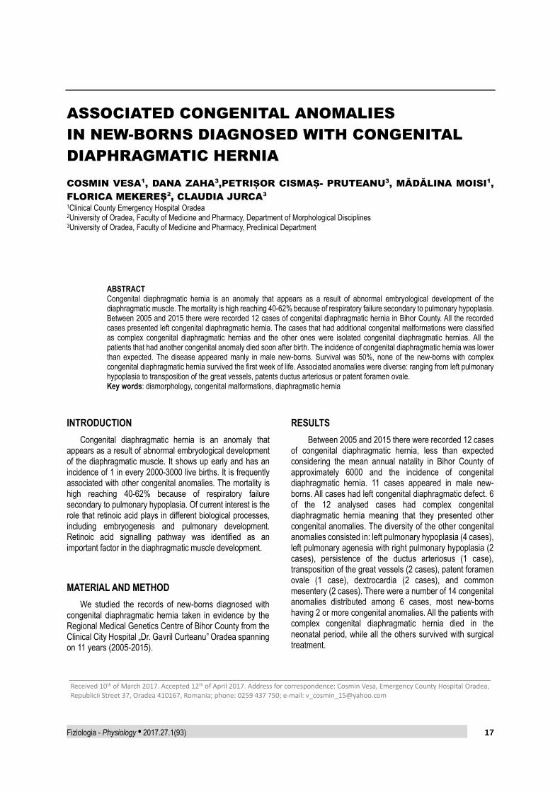
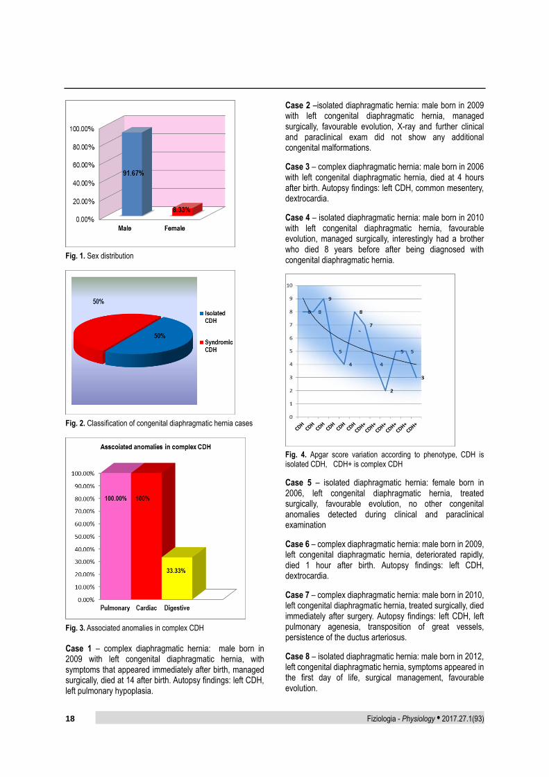
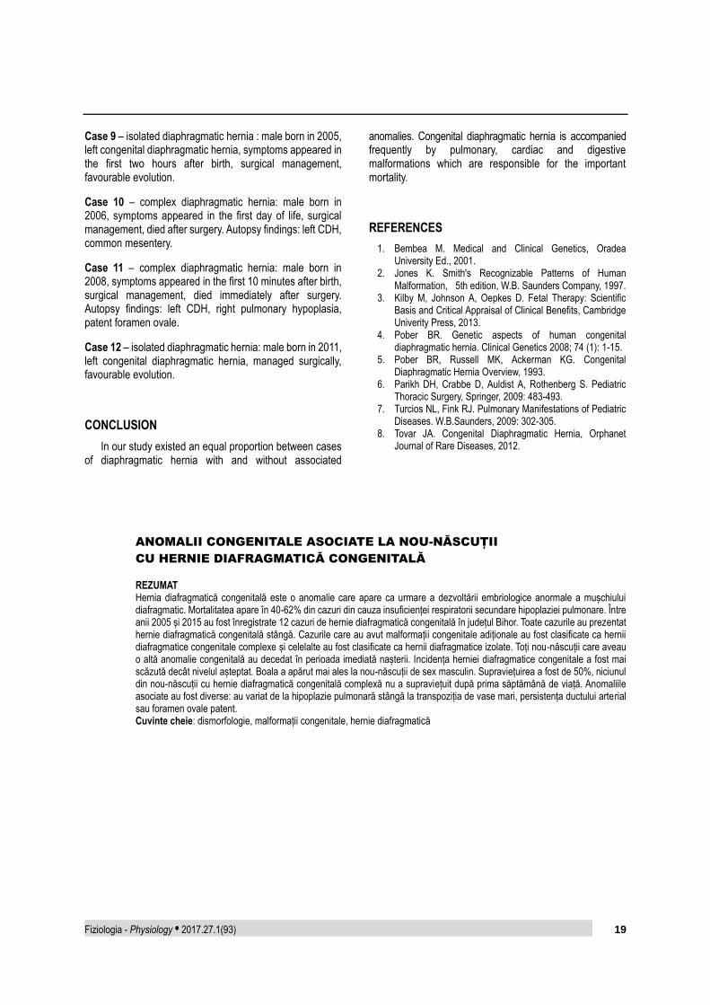
Between 2005 and 2015 there were recorded 12 cases of congenital diaphragmatic hernia, less than expected considering the mean annual natality in Bihor County of approximately 6000 and the incidence of congenital diaphragmatic hernia. 11 cases appeared in male new-borns. All cases had left congenital diaphragmatic defect. 6 of the 12 analysed cases had complex congenital diaphragmatic hernia meaning that they presented other congenital anomalies. The diversity of the other congenital anomalies consisted in: left pulmonary hypoplasia (4 cases), left pulmonary agenesia with right pulmonary hypoplasia (2 cases), persistence of the ductus arteriosus (1 case), transposition of the great vessels (2 cases), patent foramen ovale (1 case), dextrocardia (2 cases), and common mesentery (2 cases). There were a number of 14 congenital anomalies distributed among 6 cases, most new-borns having 2 or more congenital anomalies. All the patients with complex congenital diaphragmatic hernia died in the neonatal period, while all the others survived with surgical treatment.
Received 10th of March 2017. Accepted 12th of April 2017. Address for correspondence: Cosmin Vesa, Emergency County Hospital Oradea, Republicii Street 37, Oradea 410167, Romania; phone: 0259 437 750; e-mail: [email protected]
Fig. 2. Classification of congenital diaphragmatic hernia cases
Fig. 3. Associated anomalies in complex CDH
Case 1 – complex diaphragmatic hernia: male born in 2009 with left congenital diaphragmatic hernia, with symptoms that appeared immediately after birth, managed surgically, died at 14 after birth. Autopsy findings: left CDH, left pulmonary hypoplasia.
Case 2 –isolated diaphragmatic hernia: male born in 2009 with left congenital diaphragmatic hernia, managed surgically, favourable evolution, X-ray and further clinical and paraclinical exam did not show any additional congenital malformations.
Case 3 – complex diaphragmatic hernia: male born in 2006 with left congenital diaphragmatic hernia, died at 4 hours after birth. Autopsy findings: left CDH, common mesentery, dextrocardia.
Case 4 – isolated diaphragmatic hernia: male born in 2010 with left congenital diaphragmatic hernia, favourable evolution, managed surgically, interestingly had a brother who died 8 years before after being diagnosed with congenital diaphragmatic hernia.
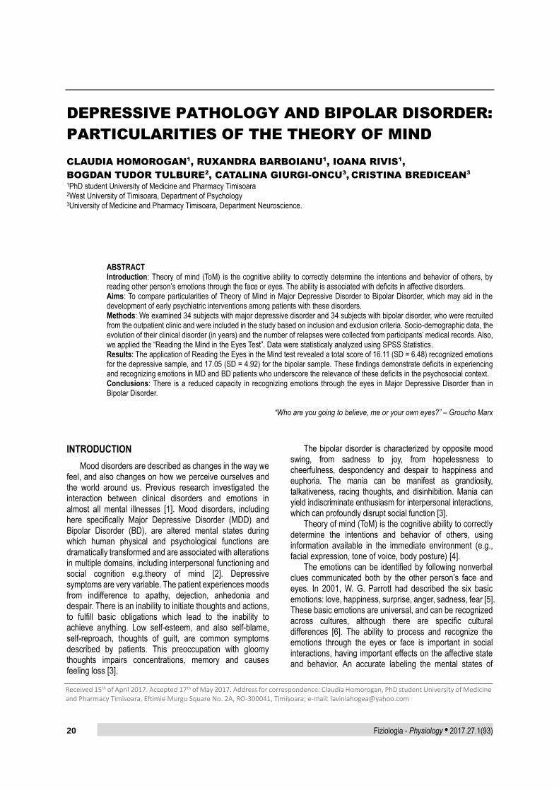
Fig. 4. Apgar score variation according to phenotype, CDH is isolated CDH, CDH+ is complex CDH
Case 5 – isolated diaphragmatic hernia: female born in 2006, left congenital diaphragmatic hernia, treated surgically, favourable evolution, no other congenital anomalies detected during clinical and paraclinical examination
Case 6 – complex diaphragmatic hernia: male born in 2009, left congenital diaphragmatic hernia, deteriorated rapidly, died 1 hour after birth. Autopsy findings: left CDH, dextrocardia.
Case 7 – complex diaphragmatic hernia: male born in 2010, left congenital diaphragmatic hernia, treated surgically, died immediately after surgery. Autopsy findings: left CDH, left pulmonary agenesia, transposition of great vessels, persistence of the ductus arteriosus.
Case 8 – isolated diaphragmatic hernia: male born in 2012, left congenital diaphragmatic hernia, symptoms appeared in the first day of life, surgical management, favourable evolution.
Fiziologia - Physiology • 2017.27.1(93) 19
Case 9 – isolated diaphragmatic hernia : male born in 2005, left congenital diaphragmatic hernia, symptoms appeared in the first two hours after birth, surgical management, favourable evolution.
Case 10 – complex diaphragmatic hernia: male born in 2006, symptoms appeared in the first day of life, surgical management, died after surgery. Autopsy findings: left CDH, common mesentery.
Case 11 – complex diaphragmatic hernia: male born in 2008, symptoms appeared in the first 10 minutes after birth, surgical management, died immediately after surgery. Autopsy findings: left CDH, right pulmonary hypoplasia, patent foramen ovale.
Case 12 – isolated diaphragmatic hernia: male born in 2011, left congenital diaphragmatic hernia, managed surgically, favourable evolution.
CONCLUSION
In our study existed an equal proportion between cases of diaphragmatic hernia with and without associated
anomalies. Congenital diaphragmatic hernia is accompanied frequently by pulmonary, cardiac and digestive malformations which are responsible for the important mortality.
REFERENCES
1. Bembea M. Medical and Clinical Genetics, Oradea University Ed., 2001.
2. Jones K. Smith's Recognizable Patterns of Human Malformation, 5th edition, W.B. Saunders Company, 1997.
3. Kilby M, Johnson A, Oepkes D. Fetal Therapy: Scientific Basis and Critical Appraisal of Clinical Benefits, Cambridge Univerity Press, 2013.
4. Pober BR. Genetic aspects of human congenital diaphragmatic hernia. Clinical Genetics 2008; 74 (1): 1-15.
5. Pober BR, Russell MK, Ackerman KG. Congenital Diaphragmatic Hernia Overview, 1993.
6. Parikh DH, Crabbe D, Auldist A, Rothenberg S. Pediatric Thoracic Surgery, Springer, 2009: 483-493.
8. Tovar JA. Congenital Diaphragmatic Hernia, Orphanet Journal of Rare Diseases, 2012.
ANOMALII CONGENITALE ASOCIATE LA NOU-NĂSCUȚII
CU HERNIE DIAFRAGMATICĂ CONGENITALĂ
REZUMAT Hernia diafragmatică congenitală este o anomalie care apare ca urmare a dezvoltării embriologice anormale a mușchiului diafragmatic. Mortalitatea apare în 40-62% din cazuri din cauza insuficienței respiratorii secundare hipoplaziei pulmonare. Între anii 2005 și 2015 au fost înregistrate 12 cazuri de hernie diafragmatică congenitală în județul Bihor. Toate cazurile au prezentat hernie diafragmatică congenitală stângă. Cazurile care au avut malformații congenitale adiționale au fost clasificate ca hernii diafragmatice congenitale complexe și celelalte au fost clasificate ca hernii diafragmatice izolate. Toți nou-născuții care aveau o altă anomalie congenitală au decedat în perioada imediată nașterii. Incidența herniei diafragmatice congenitale a fost mai scăzută decât nivelul așteptat. Boala a apărut mai ales la nou-născuții de sex masculin. Supraviețuirea a fost de 50%, niciunul din nou-născuții cu hernie diafragmatică congenitală complexă nu a supraviețuit după prima săptămână de viață. Anomaliile asociate au fost diverse: au variat de la hipoplazie pulmonară stângă la transpoziția de vase mari, persistența ductului arterial sau foramen ovale patent. Cuvinte cheie: dismorfologie, malformații congenitale, hernie diafragmatică
20 Fiziologia - Physiology • 2017.27.1(93)
DEPRESSIVE PATHOLOGY AND BIPOLAR DISORDER:
PARTICULARITIES OF THE THEORY OF MIND
CLAUDIA HOMOROGAN1
, RUXANDRA BARBOIANU1
, IOANA RIVIS1
,
BOGDAN TUDOR TULBURE2
, CATALINA GIURGI-ONCU3
,
CRISTINA BREDICEAN3
1PhD student University of Medicine and Pharmacy Timisoara 2West University of Timisoara, Department of Psychology 3University of Medicine and Pharmacy Timisoara, Department Neuroscience.
ABSTRACT Introduction: Theory of mind (ToM) is the cognitive ability to correctly determine the intentions and behavior of others, by reading other person’s emotions through the face or eyes. The ability is associated with deficits in affective disorders. Aims: To compare particularities of Theory of Mind in Major Depressive Disorder to Bipolar Disorder, which may aid in the development of early psychiatric interventions among patients with these disorders. Methods: We examined 34 subjects with major depressive disorder and 34 subjects with bipolar disorder, who were recruited from the outpatient clinic and were included in the study based on inclusion and exclusion criteria. Socio-demographic data, the evolution of their clinical disorder (in years) and the number of relapses were collected from participants’ medical records. Also, we applied the “Reading the Mind in the Eyes Test”. Data were statisticaly analyzed using SPSS Statistics. Results: The application of Reading the Eyes in the Mind test revealed a total score of 16.11 (SD = 6.48) recognized emotions for the depressive sample, and 17.05 (SD = 4.92) for the bipolar sample. These findings demonstrate deficits in experiencing and recognizing emotions in MD and BD patients who underscore the relevance of these deficits in the psychosocial context. Conclusions: There is a reduced capacity in recognizing emotions through the eyes in Major Depressive Disorder than in Bipolar Disorder.
“Who are you going to believe, me or your own eyes?” – Groucho Marx
INTRODUCTION
Mood disorders are described as changes in the way we feel, and also changes on how we perceive ourselves and the world around us. Previous research investigated the interaction between clinical disorders and emotions in almost all mental illnesses [1]. Mood disorders, including here specifically Major Depressive Disorder (MDD) and Bipolar Disorder (BD), are altered mental states during which human physical and psychological functions are dramatically transformed and are associated with alterations in multiple domains, including interpersonal functioning and social cognition e.g.theory of mind [2]. Depressive symptoms are very variable. The patient experiences moods from indifference to apathy, dejection, anhedonia and despair. There is an inability to initiate thoughts and actions, to fulfill basic obligations which lead to the inability to achieve anything. Low self-esteem, and also self-blame, self-reproach, thoughts of guilt, are common symptoms described by patients. This preoccupation with gloomy thoughts impairs concentrations, memory and causes feeling loss [3].
The bipolar disorder is characterized by opposite mood swing, from sadness to joy, from hopelessness to cheerfulness, despondency and despair to happiness and euphoria. The mania can be manifest as grandiosity, talkativeness, racing thoughts, and disinhibition. Mania can yield indiscriminate enthusiasm for interpersonal interactions, which can profoundly disrupt social function [3].
Theory of mind (ToM) is the cognitive ability to correctly determine the intentions and behavior of others, using information available in the immediate environment (e.g., facial expression, tone of voice, body posture) [4].
The emotions can be identified by following nonverbal clues communicated both by the other person’s face and eyes. In 2001, W. G. Parrott had described the six basic emotions: love, happiness, surprise, anger, sadness, fear [5]. These basic emotions are universal, and can be recognized across cultures, although there are specific cultural differences [6]. The ability to process and recognize the emotions through the eyes or face is important in social interactions, having important effects on the affective state and behavior. An accurate labeling the mental states of
Received 15th of April 2017. Accepted 17th of May 2017. Address for correspondence: Claudia Homorogan, PhD student University of Medicine and Pharmacy Timisoara, Eftimie Murgu Square No. 2A, RO-300041, Timișoara; e-mail: [email protected]
others is associated with ToM ability. Studies have showed that ToM deficits appear in schizophrenia [7], autism [8], but also in affective disorders [9,10]. Researches showed that patients with bipolar disorder displayed a pronounced deficit in the cognitive domain of ToM(4), and were found with impaired performance on theory of mind for both bipolar-depressed and bipolar-manic patients [10]. A meta-analysis of studies comparing ToM abilities in MDD and healthy controls included 18 studies comparing 613 patients with MDD and 529 healthy controls. The results have shown that MDD patients significantly underperformed healthy controls in ToM (d=0.51-0.58). ToM impairment in MDD was evident and was significantly related to severity of depressive symptoms [11]. Another study showed that reduced ToM ability in MDD was associated with poor psychosocial functioning [12].
Therefore, the aim of this study was to compare particularities of Theory of Mind in Major Depressive Disorder and Bipolar Disorder, providing evidence for social dysfunction, which may aid in the development of early psychiatric interventions among patients with these disorders.
METHODS
Participants We examined a sample of 68 individuals (N=34 with
major depressive disorder, N=34 with bipolar disorder) who have been previously hospitalized in the Psychiatry Clinic of Timisoara. Participants were recruited from the outpatient clinic, where they come for regular psychiatric assessments. All participants had a stable diagnosis of Major Depressive Disorder or Bipolar Disorder, according to ICD-10 criteria. Participants were tested while their symptoms were relatively stable, although the clinical remission was not complete. In order to take part in this study, participants had to fulfill the following inclusion criteria:
1. Current diagnostic of Major Depressive Disorder or Bipolar Disorder according to ICD-10 criteria.
2. Had at least 5 years since the diagnostic was formally registered in their medical records.
We excluded participants who: 1. Were over 65 years old; 2. Had a serious cognitive impairment or mental
retardation; 3. Had a serious organic disorder.
All Participants have been inpatients at the Psychiatry
Clinic of Timisoara and are currently monitored in the out-patient clinic.
Procedure Before the assessment, participants consented to take
part in the study by signing an informed consent. Each
participant was assessed individually. Socio-demographic data, the evolution of their clinical disorder (in years) and the number of relapses were collected from participants’ medical records. Subsequently, participants were invited to complete a self-report scale that measures their ability to recognize emotions.
Measures Reading the Mind in the Eyes Test (Baron-Cohen,
Jolliffe, Mortimore, & Robertson, 1997) consists of 36 grey-scale photos displaying one of the six different basic emotions in the area around the eyes: joy, love, surprise, anger, sadness, fear. Each photo is surrounded by four mental state terms and participants are instructed to choose the word which best describes what the person in the photo is thinking or feeling. Only one of the four items is deemed correct (as judged by consensus from an independent panel of judges in the initial psychometric study). Responses were coded as correct or incorrect, and one point is granted for each correct answer (total score rage between 0 and 36).
Data analysis Data were statisticaly analyzed using SPSS Statistics.
The two groups were compared using student t test (for their mean score) and chi square test χ2 (for the frequency of recognized emotions). Also, associations between variables was assessed using the Pearson correlation coeficient r..
Hypothesis 1. Participants with bipolar disorder will display a
higher ability of reading the eyes in the mind than those with a major depressive disorder.
2. The number of relapses will be negatively correlated with the ability of identifying emotions in both groups.
RESULTS
Sample characteristics The trial included 68 participants, divided into two equal
groups: Major Depressive Disorder (MDD), N=34 and Bipolar Disorer (BD), N=34). In both groups most participants were female (76% in MDD and 61% in BD). Average age at the moment of assessment was 55.58 years (SD = 8.37) for MDD, and 40.52 years (SD = 9.13) for BD. The average age at the onset of the disease was 43.73 years (SD = 9.38) for MDD, and 27.53 years (SD = 5.19) for BD. The age at onset of the disease and the age at the moment of assessment in both groups is significantly different: t=8.81 (p<0.00) for age at onset, and t=7.08 (p<0.00) for age at assessment. The level of education was
similar in both groups: an average of 11.85 years of school for bipolar disorder and 11.14 years for depressive disorder. Referring to professional status, in both groups, most parti-cipants were retired: 79% of depressive disorder and 68% of bipolar disorder group. In terms of marital status 71% of depressive subjects were married but only 32% of bipolar subjects were married. .The average duration of evolution for the sample with MDD was 11.44 years, and 12.7 for BD. The average numbers of relapses were 4.94 for MDD and 5.52 for BD. No differences were found between the two groups on these variables.
Reading the Eyes in the Mind test The application of Reading the Eyes in the Mind test
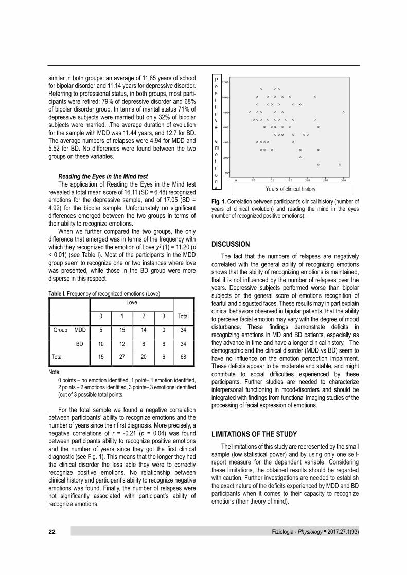
revealed a total mean score of 16.11 (SD = 6.48) recognized emotions for the depressive sample, and of 17.05 (SD = 4.92) for the bipolar sample. Unfortunately no significant differences emerged between the two groups in terms of their ability to recognize emotions.
When we further compared the two groups, the only difference that emerged was in terms of the frequency with which they recognized the emotion of Love χ2 (1) = 11.20 (p < 0.01) (see Table I). Most of the participants in the MDD group seem to recognize one or two instances where love was presented, while those in the BD group were more disperse in this respect. Table I. Frequency of recognized emotions (Love)
Love
Total 0 1 2 3
Group MDD 5 15 14 0 34
BD 10 12 6 6 34
Total 15 27 20 6 68
Note:
0 points – no emotion identified, 1 point– 1 emotion identified, 2 points – 2 emotions identified, 3 points– 3 emotions identified (out of 3 possible total points.
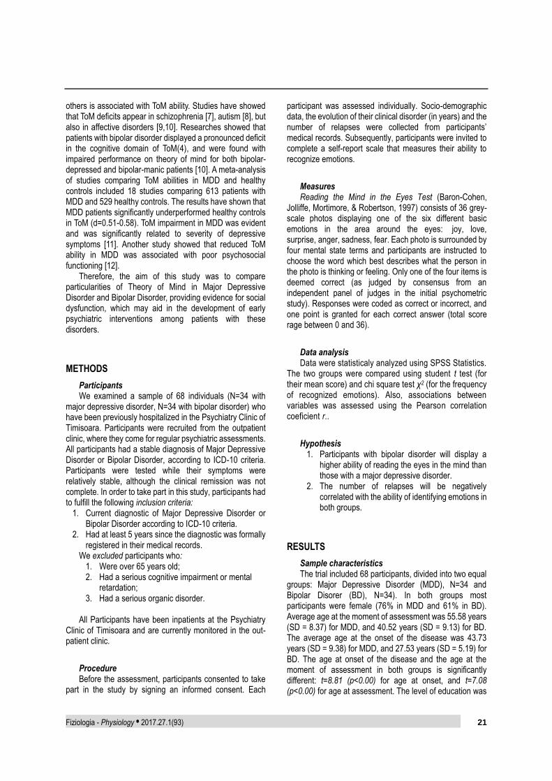
For the total sample we found a negative correlation
between participants’ ability to recognize emotions and the number of years since their first diagnosis. More precisely, a negative correlations of r = -0.21 (p = 0.04) was found between participants ability to recognize positive emotions and the number of years since they got the first clinical diagnostic (see Fig. 1). This means that the longer they had the clinical disorder the less able they were to correctly recognize positive emotions. No relationship between clinical history and participant’s ability to recognize negative emotions was found. Finally, the number of relapses were not significantly associated with participant’s ability of recognize emotions.
Fig. 1. Correlation between participant’s clinical history (number of years of clinical evolution) and reading the mind in the eyes (number of recognized positive emotions).
DISCUSSION
The fact that the numbers of relapses are negatively correlated with the general ability of recognizing emotions shows that the ability of recognizing emotions is maintained, that it is not influenced by the number of relapses over the years. Depressive subjects performed worse than bipolar subjects on the general score of emotions recognition of fearful and disgusted faces. These results may in part explain clinical behaviors observed in bipolar patients, that the ability to perceive facial emotion may vary with the degree of mood disturbance. These findings demonstrate deficits in recognizing emotions in MD and BD patients, especially as they advance in time and have a longer clinical history. The demographic and the clinical disorder (MDD vs BD) seem to have no influence on the emotion perception impairment. These deficits appear to be moderate and stable, and might contribute to social difficulties experienced by these participants. Further studies are needed to characterize interpersonal functioning in mood-disorders and should be integrated with findings from functional imaging studies of the processing of facial expression of emotions.
LIMITATIONS OF THE STUDY
The limitations of this study are represented by the small sample (low statistical power) and by using only one self-report measure for the dependent variable. Considering these limitations, the obtained results should be regarded with caution. Further investigations are needed to establish the exact nature of the deficits experienced by MDD and BD participants when it comes to their capacity to recognize emotions (their theory of mind).
Fiziologia - Physiology • 2017.27.1(93) 23
CONCLUSIONS
No differences were found between Major Depressive Disorder (MDD) and the Bipolar Disorder (BD) group regarding their ability to recognize emotions in others.
Participant’s clinical history (the number of years since their first diagnostic) were negatively associated with their general ability of recognize positive emotions. So, the longer they had the disorder the less able they were to recognize positive emotions. This decrease in their ability to recognize positive emotions as they have a longer clinical history could have detrimental effects associated with their social functioning. More well-designed studies are needed to clarify these complex relationships and shed light on the impact of affective disorders on participant’s theory of mind.
REFERENCES
1. Wood SJ, Yung AR, Pantelis C. Cognitive precursors of severe mental disorders. Cogn Neuropsychiatry. 2013; 18(1-2): 1-8.
2. Sullivan S, Lewis G, Mohr C, Herzig D, Corcoran R, Drake R, et al. The longitudinal association between social functioning and theory of mind in first-episode psychosis. Cogn Neuropsychiatry. 2014: 19(1): 58-80.
3. Oyebode F. Sims’ symptoms in the mind: Textbook of descriptive psychopathology (5th ed.). Sims’ symptoms in the mind: Textbook of descriptive psychopathology (5th ed.). 2015.
4. Montag C, Ehrlich A, Neuhaus K, Dziobek I, Heekeren HR, Heinz A, et al. Theory of mind impairments in euthymic bipolar patients. J Affect Disord 2010; 123(1-3): 264-9.
5. Parrott WG. Emotions in Social Psychology: Essential Readings. Ann Arbor: Edwards. 2001. 378 pp.
6. Sauter DA, Eisner F, Ekman P, Scott SK. Cross-cultural recognition of basic emotions through nonverbal emotional vocalizations. Proc Natl Acad Sci 2010; 107(6): 2408-12.
7. Pentaraki AD, Stefanis NC, Stahl D, Theleritis C, Toulopoulou T, Roukas D, et al. Theory of Mind as a potential trait marker of schizophrenia: A family study. Cogn Neuropsychiatry. 2012; 17(1): 64-89.
8. Sabbagh MA. Understanding orbitofrontal contributions to theory-of-mind reasoning: Implications for autism. Brain and Cognition. 2004; 55: 209-19.
9. Purcell AL, Phillips M, Gruber J. In your eyes: Does theory of mind predict impaired life functioning in bipolar disorder? J Affect Disord 2013; 151(3): 1113-9.
10. Kerr N, Dunbar RIM, Bentall RP. Theory of mind deficits in bipolar affective disorder. J Affect Disord. 2003; 73(3): 253-9.
11. Bora E, Berk M. Theory of mind in major depressive disorder: A meta-analysis. J Affect Disord. 2016; 191: 49-55.
12. Cusi AM, Nazarov A, Macqueen GM, McKinnon MC. Theory of mind deficits in patients with mild symptoms of major depressive disorder. Psychiatry Res. 2013; 210(2): 672-4.
PATOLOGIA DEPRESIVĂ ȘI TULBURĂRILE BIPOLARE:
PARTICULARITĂȚI ALE TEORIEI MINȚII
REZUMAT Introducere: Teoria minții (ToM) este capacitatea cognitive de a determina corect intențiile și comportamentul altora, prin citirea emoțiilor altor persoane la nivelul feței sau ochilor. Această abilitate este scăzută la subiecții cu tulburări afective. Scop: Compararea particularităților teoriei minții în tulburarea depresivă majoră și tulburarea bipolară, care poate ajuta la dezvoltarea unor metode intervenționale psihiatrice precoce la pacienții cu aceste afecțiuni. Metode: Am examinat 34 de subiecți cu tulburare depresivă majoră (MD) și 34 de subiecți cu tulburare bipolară (BD), care au fost recrutați din ambulator și au fost incluși în studiu pe baza criteriilor de includere și excludere. Din fișa medicală a pacienților au fost colectate date referitoare la caracteristicile socio-demografice, evoluția afecțiunii clinice (în timp) și numărul de recăderi. De asemenea, am aplicat testul “Reading the Mind in the Eyes Test”. Datele au fost analizate statistic folosind SPSS Statistics. Rezultate: Aplicarea testului ”Reading the Mind in the Eyes” a rezultat într-un scor total de 16.11 (SD = 6.48) pentru recunoașterea emoțiilor la subiecții depresivi și de 17.05 (SD = 4.92) pentru subiecții bipolari. Aceste rezultate demonstrează deficitul în trăirea și recunoașterea emoțiilor la pacienții cu MD și BD, care subliniază relevanța acestor deficite în contextul psihosocial. Concluzii: La subiecții cu depresie majoră există o capacitate mai redusă de recunoaștere a emoțiilor la nivelul ochilor decât la subiecții cu tulburare bipolară.
24 Fiziologia - Physiology • 2017.27.1(93)
PULMONARY ASPERGILLOMA IN AN
IMMUNOCOMPETENT PATIENT. A CASE REPORT
RAMONA CIOBOATA1
, ILEANA BAZAVAN1
, MARIUS GEORGESCU2
,
SAFRAN CHAUDREY1
, ALICE ELENE GAMAN3
1 Internal Medicine-Pulmonology Department, University of Medicine and Pharmacy Craiova, Romania 2 Physiology Department, University of Medicine and Pharmacy “Victor Babes”, Timisoara, Romania 3 Microbiology Department, University of Medicine and Pharmacy of Craiova, Romania;
ABSTRACT Aspergillosis is a severe pathologic condition caused by the Aspergillus organisms and is frequently seen in immunocompromised patients. Aspergillus can cause a broad spectrum of diseases in the human host, ranging from hypersensitivity reactions to direct angioinvasion. An aspergilloma is a fungus ball (mycetoma) that develops in a preexisting cavity in the lung parenchyma. Computed tomography (CT) characteristic aspect for saprophytic aspergillosis (aspergilloma) is a mass with soft-tissue attenuation within a lung cavity. The mass is typically separated from the cavity wall by airspace (“air crescent” sign) and is often associated with the thickening of the wall and adjacent pleura. The present case is an immunocompetent female patient aged 37 years, with no history of pulmonary pathology, who was admitted to our clinic with physical fatigue, fever, spontaneous micro-hemoptysis, weight loss. Following chest radiography and computed tomography the patient was diagnosed with pulmonary aspergilloma. The particularity of the case consists in the appearance of a pulmonary aspergilloma in a patient without a history of pulmonary pathology. Key words: pulmonary aspergilloma, mycetoma, hemoptysis, computed tomography
INTRODUCTION
Pulmonary aspergillosis is a rare infectious disease, caused by contamination with the Aspergillus fungi, especially the Aspergillus fumigatus and the Aspergillus Niger, comprising several locations for the pathological process, the most common of which being chronic cavitary pulmonary aspergillosis. Less common manifestations include: Aspergillus nodule and single aspergilloma [1]. Aspergillosis develops more frequently in immunocom-promised individuals. Pulmonary localization is the most common, but the spectrum of fungal infections with Aspergillus is much wider.
It appears that men are more frequently affected by this infection, the male:women ratio being 3:1. The infection is opportunistic (occurs especially when the immune system of the host is affected) and is therefore more common in immunocompromised patients. An aspergilloma is a fungus ball (mycetoma) composed of Aspergillus hyphae, fibrin, mucus, and cellular debris which develops in pre-existing lung cavities that have become colonized with Aspergillus spp [2,3]. The most common causes of pulmonary aspergilloma are tuberculosis and sarcoidosis. Less
common causes include bronchogenic cyst and bullous emphysema, while in HIV-positive patients, aspergilloma can develop in cystic areas resulting from pneumonia with Pneumocystis carinii [4].
While cavity walls are not invaded, in evolution hemoptysis, coughing and fever can manifest. The most common clinical manifestation of aspergilloma is hemoptysis, however patients may remain asymptomatic.
Patients with a single Aspergillus nodule only require antifungal therapy if it is not fully resected, but if multiple nodules are present they can benefit from antifungal treatment, while requiring a careful follow-up [1].
Surgical resection is necessary for patients with life-threatening hemoptysis. Embolization of the bronchial artery can also represent a very suitable therapeutic option, especially for patients who cannot be subjected to more sophisticated surgical interventions [5].
CASE REPORT
We present the case of an immunocompetent female patient, aged 37 years, with no history of pulmonary
Received 14th of March 2017. Accepted 10th of April 2017. Address for correspondence: Marius Georgescu, MD, PhD, Assistant Teaching Professor, Department of Functional Sciences, Physiology, Eftimie Murgu Square No. 2A, RO-300041 Timișoara; phone: 0040765527318; e-mail: [email protected]
pathology, who was admitted to our clinic for marked physical asthenia, fever, spontaneous micro-hemoptysis, weight loss.
She reported ongoing symptoms of spontaneous micro-hemoptysis and weight loss of about 5 kg over the preceding 4 weeks.
She is a current tobacco smoker with a 17-pack-years history and denied drug usage or travel abroad in the preceding year.
On examination, she was discovered to be underweight (IMC value of 20) with the following vital parameters values: a body temperature of 38.1 Celsius degrees, a heart rate of 92 beats/minutes, respiratory rate was 17 /minute and blood pressure was 130/80 mmHg.
Auscultation revealed sibilant wheeze on the upper areas of her left lung.
Routine blood examination showed normochromic normocytic anaemia (haemoglobin 10.6 g/dL, red blood cells 2.92×10⁶/mmc, and mean corpuscular volume 100), platelets at 478000/mm3 and white cell blood count was 12,700/mm3 with 61% neutrophil, 33% lymphocyte, 4% monocyte, 1% eosinophil. The sputum smear and culture for TB were negative, with no other pathogens found. TB-interferon γ release assay (TB-IGRA) result was also negative.
Chest X-ray revealed the presence of a round, non-homogeneous opacity, 3 cm in diameter, located on the upper half of her left lung.
Computerized tomography was performed and revealed an excavated lesion of irregular shape (40 mm in diameter) in the dorsal segment of the left upper lobe with solid vegetation inside, delimitated by a 3 mm thick wall and the suggestive presence of the ‘halo sign’. No signs of mediastinal adenopathy or pleural fluid are present (Figures 1, 2, 3).
Fig. 1. Computed tomography showing the mycetoma in the left upper lobe excavated lesion.
Fig. 2. Thorax computed tomogram showing excavated lesion in the dorsal segment of the left upper lobe with solid vegetation inside, delimitated by a thick wall.
Fig. 3. CT scan revealed a soft tissue opacity with ‘air crescent’ sign in the dorsal segment of the left upper lobe
DISCUSSION
Although the Aspergillus genus fungus is ubiquitous, infection with these germs is rare, the vast majority of cases being patients who are overwhelmed with antibiotics or with low immunity. It also appears that men are more frequently affected.
Aspergillosis is a serious pathological condition caused by Aspergillus species and is more commonly seen in immunocompromised patients [6].
Aspergilloma is generally diagnosed radiologicaly. The disease rarely occurs independently and is often preceded by
26 Fiziologia - Physiology • 2017.27.1(93)
pulmonary tuberculosis or other cavitating lung diseases such as bronchiectasis, necrotising pneumonia, lung neoplasia, and sarcoidosis [7, 8]. In a survey of 544 patients with post tuberculous cavities of at least 2.5 cm, with sputum negative for tuberculous bacilli, it was reported that 25% of the patients presented serum precipitins against Aspergillus, and 11% of them had radiographic evidence of aspergilloma [9].
In our case the patient was an immunocompetent female with no history of pulmonary pathology or any other pathology.
Clinical, histological and radiological manifestations are determined by the number and virulence of the Aspergillus species as well as by the patient's immune response. Although patients may remain asymptomatic, the most common clinical manifestation of pulmonary aspergilloma is hemoptysis. Other uncommon symptoms include cough, weight loss and fever [10]. Our female patient presented with spontaneous micro-hemoptysis.
Radiologically, the aspergilloma is characterized by the presence of a solid, round-oval soft tissue mass. Typically, the formation is separated from the cavity walls through a variable air-space of a semilunar shape. Occasionally, the formation can fully occupy the cavity taking its form [4].
In the case presented, the radiological examination reveals revealed presence of a round, non-homogeneous opacity, 3 cm diameter, located on the half upper part of her left lung.
Differential diagnosis is done with: the image of an abscess that does not have airway drainage or an air fluid level and appears on the chest x-ray as a round image, with a relatively well-defined contour and intensely opaque; tuberculoma [4] which is an intraparenchymatous nodular mass with dimensions ranging from 1 to 5 cm, however our patient sputum smear and culture for TB were negative as well as TB-interferon γ release assay (TB-IGRA) result; bronchiectasis [11] is defined as a permanent dilation of the bronchi, however our patient did not demonstrate any evidence of it on the CT scan; lung neoplasia, however her age in combination with the imaging investigations and clinical findings was considered unlikely; and finally sarcoidosis [12] however our female patient had no evidence of pulmonary or extrapulmonary features to sustain this diagnosis.
The appearance is more obvious in the CT examination. CT plays a significant role in differentiating the aspergilloma from other conditions like Wegener’s granulomatosis and cavitating lung carcinoma [13].
Computerized tomography in this case reveals an excavated lesion of irregular shape (40 mm in diameter) in the dorsal segment of the upper left lobe with solid vegetation inside, delimitated by a 3 mm thick wall and the suggestive presence of the ‘halo sign’.
The adequate treatment and optimal duration of therapy for mycetoma has yet to be established since this unique presentation of pulmonary aspergilloma seen in immunocompetent female patients is usually diagnosed incidentally. Treatment options include amphotericin B, voriconazole, itraconazole, caspofungin [14-16] and surgical
resection which proved to be effective in preventing recurrence of haemoptysis [17, 18].
In the present case we referred the patient to the thoracic surgery department for resection, however the patient refused the surgical intervention.
CONCLUSION
Pulmonary aspergilloma is a fungal mass that develops in pre-existing lung cavities resulting from a Koch bacillus infection, sarcoidosis, and bronchiectasis and is more commonly observed in immunocompromised patients.
The specificity of the case consists of the appearance of a pulmonary aspergilloma in an immunocompetent female with no history of pulmonary or any other pathology.
Radiology plays an essential role in the diagnosis of pulmonary aspergillosis. When the radiological examination is not conclusive, computed tomography allows the identification of the intrapulmonary space replacement process.
REFERENCES
1. Denning DW, Cadranel J, Beigelman-Aubry C, et al. Chronic pulmonary aspergillosis: rationale and clinical guidelines for diagnosis and management. Eur Respir J 2016; 47:45.
2. Judson MA, Stevens DA. The treatment of pulmonary aspergilloma. Curr Opin Investig Drugs 2001; 2:1375.
3. Denning DW, Riniotis K, Dobrashian R, Sambatakou H. Chronic cavitary and fibrosing pulmonary and pleural aspergillosis: case series, proposed nomenclature change, and review. Clin Infect Dis 2003; 37 Suppl 3:S265.
4. Franquet T, Müller NL, Giménez A, et al. Spectrum of pulmonary aspergillosis: histologic, clinical, and radiologic findings. Radio Graphics 2001, 21: 825-837.
5. Brik A, Salem AM, Kamal AR, et al. Surgical outcome of pulmonary aspergilloma. Eur J Cardiothorac Surg 2008; 34:882.
6. Kousha M, Tadi R, Soubani AO. Pulmonary aspergillosis: a clinical review. Eur Respir Rev 2011;20:156-74.
7. Hirshberg B, Biran I, Glazer M, et al. Hemoptysis: etiology, evaluation, and outcome in a tertiary referral hospital. Chest 1997;112:440-4.
8. Aspergilloma and residual tuberculous cavities-the results of a resurvey. Tubercle 1970; 51: 227.
9. Davies D. Aspergilloma and residual tuberculous cavities – the result of resurvey. Tubercle. 1970; 51: 227-45.
10. Klein DL, Gamsu G. Thoracic manifestations of aspergillosis. AJR Am. J. Roentgenol. 1980; 134: 543-552.
11. Hansell DM. Bronchiectasis. Radiol Clin North Am 1998; 36: 107-28.
12. Iannuzzi MC, Rybicki BA, Teirstein AS. et al. Engl J Med 2007; 357: 2153-65.
13. Prasad A, Agarwal K, Deepak D, Atwal SS. Pulmonary Aspergillosis: What CT can Offer Before it is too Late! JCDR. 2016; 10(4): TE01-TE05.
14. Campbell JH, Winter JH, Richardson MD, et al. Treatment of pulmonary aspergilloma with itraconazole. Thorax 1991; 46: 839.
Fiziologia - Physiology • 2017.27.1(93) 27
15. Camuset J, Nunes H, Dombret MC, et al. Treatment of chronic pulmonary aspergillosis by voriconazole in nonimmunocompromised patients. Chest 2007; 131:1435.
16. Cucchetto G, Cazzadori A, Conti M, et al. Treatment of chronic pulmonary aspergillosis with voriconazole: review of a case series. Infection 2015; 43: 277.
17. Pratap H, Dewan RK, Singh L, et al. Surgical treatment of pulmonary aspergilloma: a series of 72 cases. Indian J Chest Dis Allied Sci 2007; 49: 23.
18. Brik A, Salem AM, Kamal AR, et al. Surgical outcome of pulmonary aspergilloma. Eur J Cardiothorac Surg 2008; 34: 882.
ASPERGILOM PULMONAR LA UN PACIENT IMMUNOCOMPETENT.
PREZENTARE DE CAZ
REZUMAT Aspergiloza este o conditie patologică gravă cauzată de organismele din specia Aspergillus şi este mai frecvent observată la pacienţii imunocompromisi. Specia aspergillus poate cauza un spectru larg de afecţiuni variind de la reacţii de hipersensibilitate pană la angioinvazie. Aspergilomul este un ghem fungic (micetom) ce se dezvoltă în cavităţi pulmonare preexistente. La examenul computer tomograf aspergiloamele se caracterizează prin prezenţa unei formaţiuni solid, rotund ovalare de intensitate submediastinală. Tipic formaţiunea este separată de pereţii cavitaţii printr-un spaţiu aerian de dimensiuni variabile si de forma unei semilune. Prezentăm cazul unei paciente in varstă de 37 de ani fără antecedente pulmonare patologice şi care se prezintă in clinica noastră, pentru astenie fizica marcată, febra, microhemoptizii spontane, scădere ponderală. In urma investigatiilor imagistice in special a tomografiei computerizate se stabileşte diagnosticul de aspergilom pulmonar. Particularitatea cazului constă in apariţia unui aspergilom pulmonar la o pacientă fără antecedente pulmonare patologice. Cuvinte cheie: aspergilom pulmonar,micetom, hemoptizie, tomografie computerizată